Types of Deltoid Ligament Injuries
On this page:
- What is the deltoid ligament?
- What is a sprain of the deltoid ligament?
- How do you check the deltoid ligament?
- What are the treatment options?
- Patient story of a deltoid ligament injury
While most of us have heard of an outside ankle sprain, can you also have problems with the inside ankle ligaments? That’s called deltoid ligament injury and let’s dive into what happens and how to treat that problem without surgery.
What Is the Deltoid Ligament?

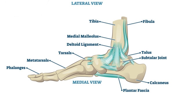
Ankle joint vector illustration. Labeled educational leg structure scheme. Physiological orthopedics explanation with isolated toe closeup. Cross section with phalanges, tibia, tarsals, ligament graph
Your ankle has ligaments on the outside and the inside. The ones on the inside are called the deltoid ligament. Think of these as the duct tape that holds your inside ankle bones together. To the left here, the ligament is shown as highlighted yellow. It has three parts, the back, middle, and front. This ligament along with others provides stability to the ankle joint including the main articulation (tibiotalar) and the joint below that (called subtalar) (1).
What Is a Sprain of the Deltoid Ligament?
A sprain is when a ligament gets stretched and injured. There are different grade sprains from lest severe (grade I) to most severe (grade III). This means that parts of the ligament have microtears.
The next type of deltoid ligament injury up from a sprain would be an incomplete tear. That’s where a part of the ligament is torn, but the other part remains intact (2). Then there’s a complete non-retracted tear, which is where the ligament is damaged all the way through, but there are fibers are still left connected.
Finally, there’s a complete retracted tear where fibers are torn in half and the two parts are snapped back like a rubber band.
How Do You Check the Deltoid Ligament?

Rumruay/Shutterstock
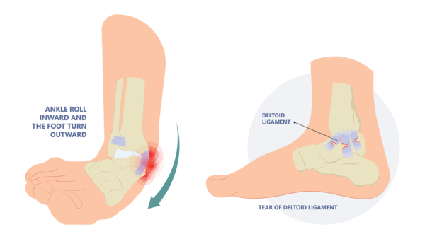
On a physical examination, a deltoid ligament injury can be checked by everting the ankle as shown here. That means taking the ankle by the heel and pulling it outward and thus stretching the inside ankle ligaments. For a patient at home, this should cause pain on the inside of the ankle. An experienced physician can also see if there’s too much motion.
There are also many different imaging options to see the extent of the deltoid ligament injury damage. These include MRI that can visualize the ligament (more on that below) or movement-based imaging techniques using ultrasound or x-ray. MRI is a type of imaging using magnetic fields that can see soft tissues like ligaments. One downside is that this is a static test, meaning if a ligament is loose, a movement-based image can show more problems than an MRI which doesn’t include movement.
Movement images of the deltoid ligament use the eversion movement as shown above plus either ultrasound or x-ray. The advantage of ultrasound is that you can directly see the ligament and how it responds (i.e. if it’s too stretchy). X-ray only looks at the bones and if they move too much, but can’t see the ligament. Both of these types of imaging can determine if the ankle is unstable. This means that the ligament is no longer protecting the joints, making them susceptible to arthritis.
What Are the Treatment Options?
Sprains are usually treated with rest and/or immobilization (for example bracing). They should heal in a few weeks. If pain or an unstable feeling continues, then the next level up for treatment of deltoid ligament injury would be injections to promote healing. Let’s review those procedures.
If the deltoid ligament is partially torn or had a complete non-retracted tear, then these injections can usually heal it:
- Please avoid steroid injections (the most commonly offered option), which may help the pain, but will inhibit healing (3).
- Prolotherapy is a solution that can prompt another inflammatory healing reaction giving your body another bite at the proverbial healing apple.
- Platelet-rich plasma (PRP) is using your body’s concentrated platelets that have healing growth factors that they release over time (4).
- Bone marrow concentrate is isolating and concentrating the stem cell fraction in your bone marrow and using that to help heal the area. Stem cells act as a general contractor for the repair response.
If the tear is a complete retracted (snapped back like a rubber band which is more unusual), then a surgical repair may be needed. This frequently involves removing the existing ligament and sacrificing a tendon to replace it by drilling holes and feeding the tendon through. Regrettably, since this is not the original equipment as Nature has designed, it will never function like a completely normal ligament.
Now let’s review actual deltoid ligament injury results.
Patient Story of a Deltoid Ligament Injury
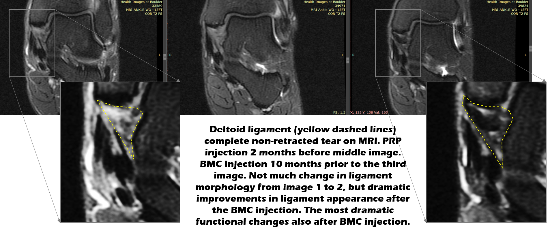
Julie is a middle-aged avid runner who was playing in the surf on a vacation and injured the inside of her ankle. Her initial MRI images are below (far left) which shows that the deltoid ligament, which is in the yellow dashed lines, is a whitish color.
A normal ligament is dark in this type of MRI image, so this indicates a full-thickness non-retracted tear. That means that the ligament looks different on the MRI all the way through, indicating damage, but it hasn’t snapped back like a rubber band. [Note, it’s critical that you have an experienced non-surgical physician take a look at the MRI films and not rely on the MRI report, as the reading radiologist may not make this distinction.]

In the images above, the middle one was taken about 2 months after a PRP injection using ultrasound guidance. Regrettably, while the patient reported some mild results, she was still having issues. In addition, the ligament is still light-colored, meaning it’s still mostly torn up. Hence, at that time, she underwent a bone marrow concentrate (BMC)injection to use her own healing stem cells.
Again, in that procedure, the BMC was precisely placed into the damaged ligament using ultrasound guidance. After that procedure, she began to feel much better and 10 months later, her new MRI to the right (3rd image) now shows a mostly normal dark appearing ligament. Her function is almost normal, but she wants to return to higher-level running, so she was back in for a second BMC treatment which she would have had earlier if not for the pandemic.
The upshot? A deltoid ligament injury may heal on its own, but if it doesn’t, there are a number of non-surgical techniques that can promote healing of the natural ligament without surgery. Julie’s deltoid ligament injury is a great example of how she was able to avoid surgery and keep her own ligament.
__________________________________________________
References
(1) Panchani PN, Chappell TM, Moore GD, et al. Anatomic study of the deltoid ligament of the ankle. Foot Ankle Int. 2014;35(9):916-921. doi:10.1177/1071100714535766
(2) Kopec TJ, Hibberd EE, Roos KG, Djoko A, Dompier TP, Kerr ZY. The Epidemiology of Deltoid Ligament Sprains in 25 National Collegiate Athletic Association Sports, 2009-2010 Through 2014-2015 Academic Years. J Athl Train. 2017;52(4):350-359. doi:10.4085/1062.6050-52.2.01
(3) Wijn SRW, Rovers MM, van Tienen TG, Hannink G. Intra-articular corticosteroid injections increase the risk of requiring knee arthroplasty. Bone Joint J. 2020 May;102-B(5):586-592. doi:10.1302/0301-620X
(4) Alsousou J, Thompson M, Hulley P, Noble A, Willett K.. The biology of platelet-rich plasma and its application in trauma and orthopaedic surgery. J Bone Joint Surg Br. 2009 Aug;91(8):987-96. doi:10.1302/0301-620X.91B8.22546

If you have questions or comments about this blog post, please email us at [email protected]
NOTE: This blog post provides general information to help the reader better understand regenerative medicine, musculoskeletal health, and related subjects. All content provided in this blog, website, or any linked materials, including text, graphics, images, patient profiles, outcomes, and information, are not intended and should not be considered or used as a substitute for medical advice, diagnosis, or treatment. Please always consult with a professional and certified healthcare provider to discuss if a treatment is right for you.