Our New C0–C1 Facet Injection Paper
This past decade, a big change has occurred in neck facet-joint treatment. The art of injecting something into a facet joint has become slowly extinct, while the procedures to perform nerve ablation of painful neck joints have ramped up. This has caused a real problem as newer regenerative-medicine techniques involving stem cells and platelet rich plasma (PRP) require the former rather than the later. Nowhere is this lack of skill more prevalent than in the upper neck. Hence, to begin addressing this problem, our group has begun to publish a series of procedural papers, this one a C0-C1 Facet Injection paper, to revive these dying procedures so that regenerative spine care can reach its full potential.
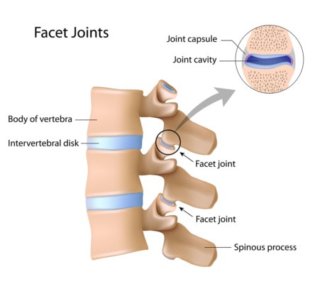
What Is a Facet Joint, and Why Is It a Big Deal?

Alila Medical Media/Shutterstock
The facets joints live between the back of the vertebrae, two at each level. They help to control motion and are about as big as a finger joint. In car-crash trauma as well as other types of injuries, they can become damaged and painful. In fact, it’s thought that damaged facet joints account for the lion’s share of why some patients develop chronic pain after a car crash.
The standard treatment for this problem for many years was injecting steroids and anesthetic into the joint. This worked to provide long-term relief in a few patients and short-term in most. In addition, since recent research has shown that injecting steroids into joints can kill cartilage, this is likely not a good idea anyway. However, due to higher reimbursement for physicians, surgery centers, and device manufacturers and better research, there’s been a distinct trend toward radiofrequency ablation (RFA).
What Is RFA?
RFA is when the doctor targets a nerve that’s taking pain signals from the damaged joint. A radiofrequency probe that looks like a thick needle is placed close to the nerve and confirmed using X-ray and electrical stimulation of the nerve. The tip of the probe heats up, burning away the nerve, hence severing the connection between the painful joint and the brain where those signals would be registered. The procedure can last for about a year (sometimes a bit less or more).
Injecting Facet Joints Is Becoming a Lost Art
Given the recent focus on RFA, it seems like as I train physicians from around the globe in various X-ray– or ultrasound-guided regenerative medicine techniques, they all recount how little experience they have with injecting the facet joints in the neck. This is doubly true of the upper-neck facet joints, where the experience levels drop to near zero. This isn’t just students but even experienced spine interventionalists. As an example, I would estimate that we have fewer than 100 physicians nationwide who have injected the top neck-facet joint (C0–C1) more than 100 times in their career.
Why Would Anyone Want to Perform a C0–C1 Facet Injection?
Many years ago, we saw a huge number of car-crash-injured patients in our Colorado practice. While the thought was that all of these patients who had headaches had injured the C2–C3 upper-neck joint, by the time patients came to us as a tertiary referral center, most had had this joint treated and still had headaches. Given that this left C1–C2 and C0–C1 as one of the several possibilities for why they still had headaches, we became expert at injecting those joints. In fact, most of those patients got better by injecting those levels, so this became a common procedure for us. As the years passed and we moved from steroids to injections like PRP and stem cells, our practice had amassed thousands of these procedures at C1–C2 and C0–C1.
Houston, We Have a Problem
For C0–C1, the traditional techniques both made this a very challenging injection. Basically, the problem was that the entry for the joint was higher than the visible joint line on X-ray, so there was no easy way to see the place where your needle needed to go to enter the joint. Over time, I invented a new way to perform the C0-C1 facet injection by estimating the entry point on the fluoroscope image. This took the procedure from a struggle to almost a sure thing to be able to document an injection in the joint. For info, see the video below:
One day a visiting physician witnessed us hit several of these joints in a row with ease and told us we should write up the new technique. Hence, our recent publication.
The upshot? Our goal is to make sure that as regenerative medicine advances, physicians can reclaim the skills they lost when RFA became “all the rage.” Now that stem cells and PRP are rapidly taking the place of destroying nerves, this new generation of physicians needs to relearn some old tricks and add some new ones as well!
If you have questions or comments about this blog post, please email us at [email protected]
NOTE: This blog post provides general information to help the reader better understand regenerative medicine, musculoskeletal health, and related subjects. All content provided in this blog, website, or any linked materials, including text, graphics, images, patient profiles, outcomes, and information, are not intended and should not be considered or used as a substitute for medical advice, diagnosis, or treatment. Please always consult with a professional and certified healthcare provider to discuss if a treatment is right for you.