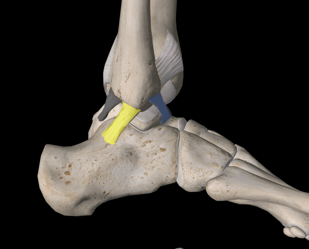
1 in 3 Unstable Ankles Has Cartilage Damage
Many people have sprained their ankles a few times in their lives. As they age, they begin to notice pain after exercise, and most ignore that pain. Now a new study shows that in 1 in 3 ankles where the ligaments are loose, there is already cartilage damage.
The Ankle Ligaments

The main ankle ligaments live on the outside and inside of the joint. The outside ligaments are more commonly sprained. These are called the ATF, CF, and PTF (Anterior Talo-fibular, Calcaneo-fibular, and Posterior Talo-fibular-see above).
These ligaments can get damaged when you roll your ankle inward. The most common type of ligament injury is an irreversible stretch of the ligament which can’t heal completely and thus causes laxity in the otherwise tight ligaments. In other words, the ligaments are no longer capable of adequately protecting the joint.
Subfailure vs. Torn Like a Rubber Band
If a ligament gets stretched and is unable to completely heal, it’s lax or loose. This isn’t usually appreciated by the patient, as once the acute pain goes away it feels fine. In addition, the ligament isn’t torn in half like a rubber band and pulled back, but instead, it’s a “sub-failure” injury. While the orthopedic surgery care system is outstanding for identifying and treating torn and retracted like a rubber band ligament tears, it almost always misses loose ligaments. That’s a big problem, as the leading cause of joint arthritis is loose ligaments.
New Research
Researchers in Amsterdam looked at multiple studies of patients with lateral ankle instability to see how many also had evidence of cartilage damage on their MRI (1). They found that 32% of ankles had a cartilage lesion, most commonly on the inside of the talus.
The Importance of Being Proactive
If you have an ankle that has been sprained a bunch of times that causes pain after exercise, then please get that ankle checked out by one of our network doctors. Unlike your orthopedic surgeon, they can use stress ultrasound to find ligaments that are damaged and stretched. An example of that type of advanced exam is above. Note that the F (Fibula) and C (Calcaneus) are pulled much farther apart in the left ultrasound-which is the unstable side. The ultrasound image on the right shows less movement and is stable.
If the ankle was unstable, what would the treatment look like? The doctor uses ultrasound guidance to carefully place high-dose platelet-rich plasma into the ligaments discussed above. This usually tightens these ligaments and provides stability in 4-6 weeks or a bit longer if you’re older.
The upshot? Loose ankle ligaments lead to ankle arthritis. So if your ankle is painful or swollen after exercise, get it checked!
__________________________________________________
References:
- Wijnhoud EJ, Rikken QGH, Dahmen J, Sierevelt IN, Stufkens SAS, Kerkhoffs GMMJ. One in Three Patients With Chronic Lateral Ankle Instability Has a Cartilage Lesion. The American Journal of Sports Medicine. April 2022. doi:10.1177/03635465221084365

NOTE: This blog post provides general information to help the reader better understand regenerative medicine, musculoskeletal health, and related subjects. All content provided in this blog, website, or any linked materials, including text, graphics, images, patient profiles, outcomes, and information, are not intended and should not be considered or used as a substitute for medical advice, diagnosis, or treatment. Please always consult with a professional and certified healthcare provider to discuss if a treatment is right for you.
