Learning How Best to Inject the CCJ Ligaments: Our CCJ Instability Trial Begins…
Last week, we began the world’s first randomized controlled placebo trial to treat craniocervical junction (CCJ) instability. While our clinic has been treating these patients for more than two decades, these past few years, we have developed a new injection technique to finally begin addressing the problem in ways that existing care can not. Let me explain.
What Is CCJ Instability?
The area where your head meets the spine is called the CCJ, which is short for the craniocervical junction. There are many strong ligaments here that help to hold your head on, which include the alar, transverse, accessory, apical dens, and others. These ligaments can be injured or become loose. The two most common causes are trauma (like a car crash) and a congenital tissue abnormality called Ehler-Danlos syndrome (EDS).
When these ligaments allow too much motion, the patient can have all sorts of symptoms including headache, muscle tightness, dizziness, and poor cognition, to name a few. In addition, they also tend to be quite disabled, and nothing really helps long-term. Physical therapy usually causes the symptoms to get worse, so they soon learn to avoid that treatment. In fact, for many, the only thing that usually helps short-term is an upper-cervical manipulation.
What Treatments Are Currently Available?
Many of these patients end up getting told that they need an upper-cervical fusion, and some actually get one. This is a huge and risky surgical procedure where the head is bolted to the upper-neck bones. This procedure has far more side effects than the average neck fusion, and as a result, there are few places that actually perform it. There are, however, a few areas of excellence that specialize in CCJ fusion.
Alternative therapies abound. A popular one is prolotherapy, which involves injections of a chemical irritant into the ligaments to increase their strength. The biggest issue is that most of the important ligaments that hold the head on can’t be accessed by this procedure, which focuses on the ligaments that can be injected from the back of the spine. In addition, prolotherapy has the disadvantage of many blind injections. Meaning that the proponents of this technique tend not to use imaging guidance or use it not to guide the needle but to confirm the position after it’s been placed. The latter is a little shooting practice where you need to improve your aim of the target after you’ve pulled the trigger.
A New Approach Emerges
A few years ago, I began to theorize that you could inject the deep CCJ ligaments (alar, transverse, and accessory) another way. This would be coming from the front and passing the needle through a small opening between the C1 and C2 neck bones. While this would involve injecting through the back of the throat, this would be the safest route to get an agent that could facilitate healing into these damaged ligaments.
For more info on this procedure (when I had only injected 50 or so patients), see the video below. Given that I’m now up to about 80 patients in late spring of 2018, I’ve learned a few things since then.
Learning How to Inject This Area
While for many areas of medicine that involve precise X-ray– or ultrasound-guided procedures, there are textbooks that you can read or research papers that can give you insight into the right or wrong ways to approach that procedure, nothing like that exists for this procedure. While this posterior oropharynx (back of the throat) approach has been used rarely to inject the C1 and C2 bones with bone cement when there is cancer destroying the area, nobody has ever used contrast injections to deliver healing substances to these damaged ligaments. This means that with every procedure, we were and still are learning how best to inject this area.
First, we had simple concerns about how best to get access to this area. While that may seem like a trivial problem, it’s actually not. Mouthpieces that keep the tongue down and out of the way aren’t designed to be invisible in an X-ray procedure. Once we found one that we could use, then the next issue was the best type of anesthesia and being able to see where you were injecting. For the latter, we use a flexible endoscope. We also finally had to design a 3D-printed mouthpiece for our own use, as the commercial off-the-shelf one we began to use was suboptimal.
Finally, once all of the issues surrounding how best to access this area were addressed, the next step was the actual injection procedure. At which angle should the needle be placed? What are the injection targets? What does radiographic contrast look like when injected into these ligaments accurately and when they are missed? Below is an example of a recent discovery that highlights that process.
What the Heck Is This “C” Shaped Structure?
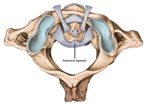
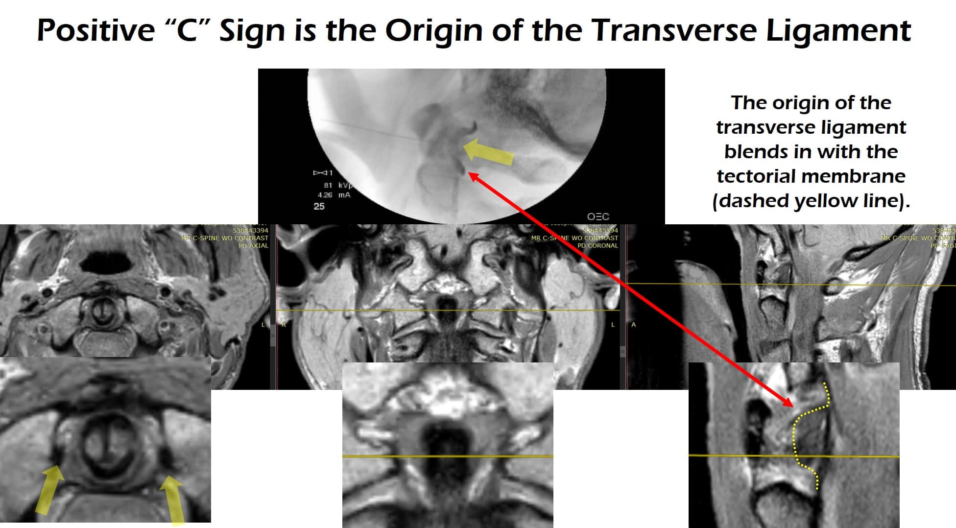
Once our new 3D-printed mouthpiece was in use, this allowed us to have more choice of injection angles. When we used these improved angles to get to these ligaments, we began to see a curious “C” shape appear on some injections. This was an area where the radiographic contrast would pool. Was this the bursa that’s in this area? Was it a blood vessel? Once we had a chance to rule in or out various things and spend time reviewing various research papers on the anatomy of this area, we realized that this was where the transverse ligament attached to the inner wall of the atlas. So our “C” was, in fact, a good thing, as it was confirmation that we were accurately injecting this ligament. See below for an image of the transverse ligament and where the injection occurred (where the ligament attaches to the inner wall):

Stihii/Shutterstock
These are the images (fluoroscopy above and the patient’s MRI below):
This type of learning is critical for any new procedure.
Our New RCT Treats Its First Patient
Like any new procedure, we need to “put our money where our mouth is.” While we have seen excellent results with this new CCJ injection procedure in a patient population that rarely gets much long-term benefit from anything, we now need to prove that it works. To that, we’ve spent the last year getting ready for a placebo-controlled randomized controlled trial. The CCJ Instability Trial began this past week.
In the new study, the patient will be randomized to either get the injection or a poke in the back of the throat without the procedure being performed. The patient won’t know which he or she received until six months and then will be unblinded. At that point, if the patient had the placebo procedure, he or she can opt to get the actual procedure.
The upshot? It’s an amazing opportunity for a physician to get to pioneer new procedures and to see patients who don’t improve with anything report life-changing results. Like any good physician, I learn more things with every procedure I perform. However, it’s now time for us to prove with an RCT that the results we’re seeing aren’t the placebo effect. We look forward to that data.

NOTE: This blog post provides general information to help the reader better understand regenerative medicine, musculoskeletal health, and related subjects. All content provided in this blog, website, or any linked materials, including text, graphics, images, patient profiles, outcomes, and information, are not intended and should not be considered or used as a substitute for medical advice, diagnosis, or treatment. Please always consult with a professional and certified healthcare provider to discuss if a treatment is right for you.

