Patients often get a shiny new shoulder MRI CD and pop it into their computer before they have the report from the radiologist. For those situations, I have a video on how to read a shoulder MRI, provided at the bottom of this page.
However, once you have the Shoulder MRI Report in hand, the next step is usually a series of Google searches in an attempt to decipher all of the medical jargon and anatomical references. This is a comprehensive guide to those obscure terms and what treatments are usually effective. Basically, how to read a shoulder MRI report.
The Key Parts of the Shoulder
First, realize that the shoulder is broken up into a few key parts:
Rotator cuff – This is made up of the supraspinatus, infraspinatus, subscapularis, and teres muscles and tendons.
Glenohumeral (GH)/AC joint – The GH joint is the main shoulder joint. AC Joint is the joint between the collar bone (clavicle) and shoulder blade (scapula).
Labrum/biceps – The labrum is the lip on the socket of the ball and socket shoulder joint. The biceps tendon attaches at the top of the lip of the main shoulder socket.
The Bursas – These are lubricating sacs around the shoulder that help ease movement of tendons past one another or another structure.
Focus on the Impression
The last part of the report with the conclusions of the radiologist is called the “Impression”. This is where you should focus your attention. One or more of the four areas above will be commented on if there’s an abnormality. I’ll break down what you might read here by each of these areas:
Rotator cuff
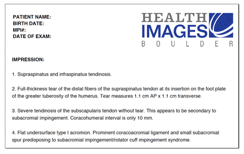
The radiologist will comment here if one of the key muscles and/or tendons is injured or damaged (supraspinatus, infraspinatus, subscapularis, and teres muscles and tendons). Issues in the supraspinatus are the most common. These are the terms that are commonly used:
A. Tendinosis/Tendinopathy/Tendinitis – For all practical purposes these all really mean about the same thing – a pissed off, degenerated, and/or swollen tendon.
B. Partial, partial thickness, or incomplete tear – This is what it sounds like, a tear in the tendon or muscle that doesn’t go all the way through.
C. Complete or full thickness tear – This is again what it sounds like, a tear all the way through the tendon. Be a bit careful here as this only means that the tear is through the tendon in one part. Look for other key terms in the body of the report or in the impression like “retracted” or “massive” because these point to a more severe full thickness tear. If those terms aren’t present, then your tear is likely smaller.
The good news is that an irritated tendon or one that’s partially torn is usually easy to help with physical therapy and/or a simple injection. Stay away from cortisone or steroid shots, as these will only weaken the shoulder tendon. In addition, any injection into this area of the shoulder should only be performed with ultrasound guidance, so stay clear of doctors who perform blind and/or unguided injections, as they may be injecting into the wrong spot. Finally, a platelet rich plasma shot is usually a good option for this type of issue, but only if performed under precise ultrasound guidance.
A. Osteoarthritis (OA) – Mild, moderate, severe – this means lost cartilage.
B. Osteophytes – This means bone spurs. These can also be called spurs or spurring. These are outgrowths of the bone caused by instability.
c. Effusion – This means swelling with fluid in the joint.
For mild OA and sometimes moderate, platelet rich plasma (PRP) usually works well to reduce pain and swelling. Also realize that arthritis may be caused by instability in the joint, due to loose ligaments. Instability usually responds well to ligament tightening injections. OA patients are often offered steroid shots as an option to relieve pain. These shots can chew up cartilage in the joint and make things worse in the long run, so they should be avoided.
The labrum is the lip around the socket of the main shoulder joint.
SLAP Tear, biceps tendon anchor tear – These are sometimes classified as types 1-4. Types 1 and 2 are less severe. Type 1 is usually treated with physical therapy. Type 2 can often be treated with a precise ultrasound guided platelet rich plasma injection. Be aware that this is a high skill procedure with only about 100-200 U.S. physicians capable of performing with a high degree of accuracy. Surgery is usually not needed for these types of labral tears, but is frequently recommended. Type 3 may be responsive to stem cell injections with the same caveats as above. Type 4 may need surgery. However, realize that surgery results for SLAP tears aren’t great.
Labral tear – This lip structure is torn. A specific type of labral tear is known as a Bankart lesion or tear. Many times radiologists will locate the tear on the socket by using a clock face naming system where 12 o’clock is at the top of the socket, 3 pm is the front, 6 pm is the bottom, and 9 pm is the back. Most smaller tears can be treated with precise platelet rich plasma injections.
Labral fraying or fraying of the labrum – This is the same as type 1 above and is usually something treated with physical therapy. PRP injections can also help.
Biceps tendon tear – These are partial or complete tears, similar to the types of rotator cuff tears discussed above with similar recommendations for treatment for each type. If you have a massive biceps tendon tear you will know it without an MRI as some or all the biceps muscle will fall downward, making you look like “Popeye”.
Bursas
These are the lubricating sacs around the shoulder that allow normal motion of tendons as they cross each other and bony areas. They can swell when they get irritated.
Effusion or fluid in the bursa – this means swelling in the bursa and is classified as mild, moderate, or severe. Realize that swelling in these areas is usually due to problems with bio mechanics and aren’t issues themselves. So this is a symptom of a bigger problem.
Impingement, sub-acromial impingement, rotator cuff impingement, type 1, 2, or 3 acromion – This means that the rotator cuff is usually being pressed on or compressed by either the way you move or a bone spur. The acromion is a natural part of the shoulder blade that can place downward pressure on the rotator cuff. The higher the type number, the more downward pressure. For example a type 3 places more pressure than than a type 1.
The upshot? Reading a shoulder MRI report and understanding what it means can be empowering because it means the patient is armed with knowledge. Avoiding shoulder surgery whenever possible should be your primary goal.
Ready to learn how to tie this information together with your MRI CD? Watch the video below.
The shoulder is an incredibly complex joint and when you put it into 3D space through the power of MRI imaging, it can be pretty difficult to figure out where all of the components described above are located. This video will help you understand what you’re looking at when you go exploring your Shoulder MRI CD. Request a Regenexx Appointment
This blog post provides general information to help the reader better understand regenerative medicine, musculoskeletal health, and related subjects. All content provided in this blog, website, or any linked materials, including text, graphics, images, patient profiles, outcomes, and information, are not intended and should not be considered or used as a substitute for medical advice, diagnosis, or treatment. Please always consult with a professional and certified healthcare provider to discuss if a treatment is right for you.
Get health and wellness information from a trusted source.
By submitting the form, you are agreeing that you read and consent to our Privacy Policy. We may also contact you via email, phone, and other electronic means to communicate information about our products and services. We do not sell, or share your information to third party vendors.
This is awesome information, Doc. I am 36, been lifting weights for 20 years, had just gotten my bench up to 500 pounds, and then my shoulders started acting up. My doc says I have osteoarthritis in a few joints, but my right shoulder had gotten so bad that I haven’t lifted in about 2 months–I THINK I tore something in there. I just had my MRI in a big MRI tube because I am too wide to fit in a regular one. I have the CD and am anxiously awaiting the report so I can know what needs to be done to get me back in the gym! I will be consulting this article again after seeing the report I get. Thank you!
so, if my doctor has been doing “blind” steroid injections to my right shoulder off and on for the last 5 years, he is just helping me along to the operating table? My shoulder locks up randomly, previous MRI’s show arthritis, cysts, spurs and the like but never any tears. he does one or sometimes two injections and I do my PT exercises religiously and I don’t end up back in his office for a year or more. then I reach the wrong way and pop – boom dinosaur arm! off for an injection and I’m good to go – or am I doing more harm than good getting the wrong kind of shot and how to I tell my ortho shoulder specialist um, hey I think you are doing that wrong?
Karen, Unfortunately, steroid injections are a net negative as they are lethal to the local stem cells which are your body’s repairmen in your joints, break down cartilage, and can injure tendons. Unfortunately as well, they remain a mainstay of orthopedic care as they are covered by insurance and despite the well documented longterm damage, they make patients feel better in the short term. Please see: https://regenexx.com/blog/bad-news-shoulder-steroid-shots/ and https://regenexx.com/steroid-injection-risks/
Thanks for this – getting MRI tonight – have a confirmed Glenoid 20% dominant arm break of 7wks old (from a serious fall) 1st Orthopedic took non surgical approach – sling through this week. Pain still acute and cannot take arm away from/above, behind body w/out acute pain. Sought 2nd opinion – leads to this 1st MRI tonight as he is suspecting possible RC tear as well – 1st Ortho ruled out by in ofc. ultrasound only. Do you agree that that this is a good route? I cannot work as a Sign Language Interpreter (I am Nationally Certified and it is my full time profession) without the full use and range of motion of my arm which I am unsure the 1st Ortho really grasped. Agreed on this route of the 2nd Ortho? Thank you!
We’d need more information like an MRI, medical history, etc, which are submitted as part of the Candidacy process. Ruling out a Rotator Cuff Tear is a good idea, although, it could be less obvious things as well. What would not be a good idea would be Rotator Cuff surgery. If you’d like us to weigh in, please submit the Candidate form. Please see: https://regenexx.com/the-regenexx-procedures/shoulder-surgery-alternative/
My disc has my neck MRI images and MRI transcribed report. The disc is supposed to have the transcribed report for my left shoulder with and without contrast too, but there is no dictated / transcribed report – only a blank header. Is the news that bad, they didn’t want me nosing through the disc before my doctor could talk to me? Or was it Friday, they had too much going on and forgot. What’s the common practice here? Princess Valium just wore off from the MRI study – I’ll be up all night now.
Incidentally, I was a medical transcriptionist for many years and understood the MRI report of my neck – standard jargon.
I can’t wait to compare my shoulder report to this site. Saved to my favorites – thank-you!
Laura, No way to know. We don’t use the report. Our Doctors read the MRI images directly as radiologists are looking for completely different things than we are looking for.
Hi there. I got these results today from my MRI 4 days ago. (I had a torn rotator cuff repair and bicep tenodesis 3 years ago on the left shoulder – successful.) Is this similar? (Rheumatoid arthritis is also a problem.)
” Patient I believe has a full-thickness tear of the supraspinatus tendon with a gap in the tendon measuring approximately 2.5 cm. The infraspinatus tendon appears partially torn. Subscapularis tendon may be partially torn on its superior aspect. The biceps tendon demonstrates abnormal signal in its distal portion probably representing some tendinopathy. Superior labrum demonstrates no bony abnormality.
IMPRESSION: Full-thickness rotator cuff tear with gap in the supraspinatus tendon. Probable abnormal biceps tendon.
Jill, Not sure what you’re referring to when you ask if this is similar, as there are many different issues discussed on the “Read Your Own Shoulder MRI” Blog. Current MRI not unusual for a patient having had a rotator cuff surgery and tenodesis.
Just wondering if the impression posted above for my right shoulder is similar to the situation from a few years ago on my left shoulder – rotator cuff repair and bicep tenodesis. Sorry – I should have made that more clear. Different techs and different medical offices doing the MRIs use different language – not the same wording for the right shoulder (above) as it was for my left shoulder (now fixed).
Jill, we take looking at MRIs very seriously as part of the candidacy process. Hence, I would need to review the films to see if you’re a candidate for what we do. We also have many excellent physicians on the network throughout the country who could do the same.
My MRI findings state that tendinopathy of the supraspinatus and infraspinatus tendon. No fluid-filled rotator cuff tear. What does this mean do I have a messed up rotator cuff?
Does a shoulder MRI include imaging of the SC joint? I have swelling over my SC joint and it hurts to touch. I have excruciating shoulder pain and neck pain. My doctor has ordered an MRI of my shoulder but I wasn’t sure if it will show the SC joint.
Hi Liz, No, a routine shoulder MRI doesn’t include the SC joint. Neck and shoulder pain are often related. Was this due to an injury, or did it just develop over time?
Reading about the cortisone shot scared me, in the beginning of my evaluation the dr. suggested me the shot because it will “ease” the pain cause what he thought i have ( just tendinitis) after the shot ( w/ ultrasound) at night i couldn’t sleep because i felt uncomfortable with a little pain on an area opposite to the shot and it felt more tender than before … so it didn’t help me at all. I continued with PT for 4weeks with a minor improvement so the Dr decided to check throughly with an MRI showing :
Tendinosis of the infraspinatus and supraspinatus tendons.
Moderate AC joint arthrosis. Downsloping acromium with a 10 mm acromial spur. Minimal subacromial bursitis.
Posterior inferior labral tear extending from the 9:00 to the 6:00 position.
Thanks for let me understand a bit of what i have and be prepared for my follow up appointment with the dr. The only thing i didn’t understand on the report was this: Findings consistent with capsulitis ( what does this mean?)
I just had my MRI report and it found “the thickening of synovium and capsule in the axillary recess”, There is also abnormal soft tissue thickening within the rotator interval encasing the biceps anchor. Overall findings are compatible with adhesive capsulitis. Does it need an operation?
I had an MRI of my left shoulder in April 2020 showing some bursitis, tendinitis, bone spur and rotator cuff fraying. My dr suggested pt for 5 weeks My motion got better for about 2 weeks, then pain in arm started up again with limited motion. My most recent pain is now in the right arm for 5 months now. Pain near clavicle and running down arm. More pain on inner bicep. Most pain during the night and tightness in morning. If I put pressure on my shoulder, pain is mild to severe. My arms are usually cold and now I’m having pain in back of neck radiating more towards right arm, but left arm has been painful also for several months now. Will see orthopedist soon. What could be wrong?
Chris Centeno, MD is a specialist in regenerative medicine and the new field of Interventional Orthopedics. Centeno pioneered orthopedic stem cell procedures in 2005 and is responsible for a large amount of the published research on stem cell use for orthopedic applications.
Learn About the #1 Stem Cell & Platelet Procedures for Treating Shoulder Arthritis & Rotator Cuff Tears - Regenexx
Get Blog Updates by Email
Get fresh updates and insights from Regenexx delivered straight to your inbox.
Find a Regenexx Provider
Regenexx is a world-wide network of specially trained physicians providing the world's most advanced, research-driven, regenerative-medicine treatments in North America, Europe, the Middle East, Asia and Australia.
Regenerative procedures are commonly used to treat musculoskelatal trauma, overuse injuries, and degenerative issues, including failed surgeries.
Shoulder
Many Shoulder and Rotator Cuff injuries are good candidates for regenerative treatments. Before considering shoulder arthroscopy or shoulder replacement, consider an evaluation of your condition with a regenerative treatment specialist.
Many spine injuries and degenerative conditions are good candidates for regenerative treatments and there are a number of studies showing promising results in treating a wide range of spine problems. Spine surgery should be a last resort for anyone, due to the cascade of negative effects it can have on the areas surrounding the surgery. And epidural steroid injections are problematic due to their long-term negative impact on bone density.
Knees are the target of many common sports injuries. Sadly, they are also the target of a number of surgeries that research has frequently shown to be ineffective or minimally effective. Knee arthritis can also be a common cause for aging athletes to abandon the sports and activities they love. Regenerative procedures can be used to treat a wide range of knee injuries and conditions. They can even be used to reduce pain and delay knee replacement for more severe arthritis.
Many spine injuries and degenerative conditions are good candidates for regenerative treatments and there are a number of studies showing promising results in treating a wide range of spine problems. Spine surgery should be a last resort for anyone, due to the cascade of negative effects it can have on the areas surrounding the surgery. And epidural steroid injections are problematic due to their long-term negative impact on bone density.
Hand and wrist injuries and arthritis, carpal tunnel syndrome, and conditions relating to overuse of the thumb, are good candidates for regenerative treatments. Before considering surgery, consider an evaluation of your condition with a regenerative treatment specialist.
Hand and Wrist Arthritis
Carpal Tunnel Syndrome
Trigger Finger
Thumb Arthritis (Basal Joint, CMC, Gamer’s Thumb, Texting Thumb)
Hip injuries and degenerative conditions become more common with age. Do to the nature of the joint, it’s not quite as easy to injure as a knee, but it can take a beating and pain often develops over time. Whether a hip condition is acute or degenerative, regenerative procedures can help reduce pain and may help heal injured tissue, without the complications of invasive surgical hip procedures.
Foot and ankle injuries are common in athletes. These injuries can often benefit from non-surgical regenerative treatments. Before considering surgery, consider an evaluation of your condition with a regenerative treatment specialist.
DISCLAIMER: The procedures and claims made about particular procedures on or through this site have not been evaluated or approved by the FDA. All statements, opinions and references provided by GetRegenerative.com are for informational purposes only. They do not constitute an endorsement of any medical provider nor guarantee the efficacy of the treatments provided. Get Regenerative does not diagnose or treat medical conditions via this website or via telephone. This site disclaims any liability for damages or negative consequences from treatments provided by the physicians listed herein to any person referencing GetRegenerative.com.
*DISCLAIMER: Like all medical procedures, Regenexx® Procedures have a success and failure rate. Patient reviews and testimonials on this site should not be interpreted as a statement on the effectiveness of our treatments for anyone else.
Providers listed on the Regenexx website are for informational purposes only and are not a recommendation from Regenexx for a specific provider or a guarantee of the outcome of any treatment you receive.
This is awesome information, Doc. I am 36, been lifting weights for 20 years, had just gotten my bench up to 500 pounds, and then my shoulders started acting up. My doc says I have osteoarthritis in a few joints, but my right shoulder had gotten so bad that I haven’t lifted in about 2 months–I THINK I tore something in there. I just had my MRI in a big MRI tube because I am too wide to fit in a regular one. I have the CD and am anxiously awaiting the report so I can know what needs to be done to get me back in the gym! I will be consulting this article again after seeing the report I get. Thank you!
Bigg Scott,
Sounds like a plan. As well as MRI which is a static image, a Stress Shoulder Ultrasound can give significantly more diagnostic information. Please see: https://regenexx.com/blog/shoulder-instability-recovery-time/
so, if my doctor has been doing “blind” steroid injections to my right shoulder off and on for the last 5 years, he is just helping me along to the operating table? My shoulder locks up randomly, previous MRI’s show arthritis, cysts, spurs and the like but never any tears. he does one or sometimes two injections and I do my PT exercises religiously and I don’t end up back in his office for a year or more. then I reach the wrong way and pop – boom dinosaur arm! off for an injection and I’m good to go – or am I doing more harm than good getting the wrong kind of shot and how to I tell my ortho shoulder specialist um, hey I think you are doing that wrong?
Karen,
Unfortunately, steroid injections are a net negative as they are lethal to the local stem cells which are your body’s repairmen in your joints, break down cartilage, and can injure tendons. Unfortunately as well, they remain a mainstay of orthopedic care as they are covered by insurance and despite the well documented longterm damage, they make patients feel better in the short term. Please see: https://regenexx.com/blog/bad-news-shoulder-steroid-shots/ and https://regenexx.com/steroid-injection-risks/
Thank you. Really appreciate this explanation.
Gald you enjoyed it Cathy!
Thanks for this – getting MRI tonight – have a confirmed Glenoid 20% dominant arm break of 7wks old (from a serious fall) 1st Orthopedic took non surgical approach – sling through this week. Pain still acute and cannot take arm away from/above, behind body w/out acute pain. Sought 2nd opinion – leads to this 1st MRI tonight as he is suspecting possible RC tear as well – 1st Ortho ruled out by in ofc. ultrasound only. Do you agree that that this is a good route? I cannot work as a Sign Language Interpreter (I am Nationally Certified and it is my full time profession) without the full use and range of motion of my arm which I am unsure the 1st Ortho really grasped. Agreed on this route of the 2nd Ortho?
Thank you!
Janet,
We’d need more information like an MRI, medical history, etc, which are submitted as part of the Candidacy process. Ruling out a Rotator Cuff Tear is a good idea, although, it could be less obvious things as well. What would not be a good idea would be Rotator Cuff surgery. If you’d like us to weigh in, please submit the Candidate form. Please see: https://regenexx.com/the-regenexx-procedures/shoulder-surgery-alternative/
My disc has my neck MRI images and MRI transcribed report.
The disc is supposed to have the transcribed report for my left shoulder with and without contrast too, but there is no dictated / transcribed report – only a blank header.
Is the news that bad, they didn’t want me nosing through the disc before my doctor could talk to me? Or was it Friday, they had too much going on and forgot.
What’s the common practice here? Princess Valium just wore off from the MRI study – I’ll be up all night now.
Incidentally, I was a medical transcriptionist for many years and understood the MRI report of my neck – standard jargon.
I can’t wait to compare my shoulder report to this site. Saved to my favorites – thank-you!
Laura,
No way to know. We don’t use the report. Our Doctors read the MRI images directly as radiologists are looking for completely different things than we are looking for.
Hi there. I got these results today from my MRI 4 days ago. (I had a torn rotator cuff repair and bicep tenodesis 3 years ago on the left shoulder – successful.) Is this similar? (Rheumatoid arthritis is also a problem.)
”
Patient I believe has a full-thickness tear of the supraspinatus tendon with a gap in the tendon measuring approximately 2.5 cm. The infraspinatus tendon appears partially torn. Subscapularis tendon may be partially torn on its superior aspect. The biceps tendon demonstrates abnormal signal in its distal portion probably representing some tendinopathy. Superior labrum demonstrates no bony abnormality.
IMPRESSION: Full-thickness rotator cuff tear with gap in the supraspinatus tendon. Probable abnormal biceps tendon.
Jill,
Not sure what you’re referring to when you ask if this is similar, as there are many different issues discussed on the “Read Your Own Shoulder MRI” Blog. Current MRI not unusual for a patient having had a rotator cuff surgery and tenodesis.
Just wondering if the impression posted above for my right shoulder is similar to the situation from a few years ago on my left shoulder – rotator cuff repair and bicep tenodesis. Sorry – I should have made that more clear. Different techs and different medical offices doing the MRIs use different language – not the same wording for the right shoulder (above) as it was for my left shoulder (now fixed).
Jill, we take looking at MRIs very seriously as part of the candidacy process. Hence, I would need to review the films to see if you’re a candidate for what we do. We also have many excellent physicians on the network throughout the country who could do the same.
My MRI findings state that tendinopathy of the supraspinatus and infraspinatus tendon. No fluid-filled rotator cuff tear. What does this mean do I have a messed up rotator cuff?
Crystal,
We don’t use reports, we’d need to see the MRI images themselves in a Candidacy evaluation, but if it hasn’t healed on it’s own, would likely benefit from treatment. This tear din’t show up on the MRI: https://regenexx.com/blog/watch-advanced-image-guided-injection-shoulder-step-procedure-suite-dr-schultz/
This would mean that your tendons have small micro tears and are “beat up”, but not completely torn.
I got my MRI report what does a dehiscent ac joint mean?
That’s not a common descriptor used in reports about AC joints, but it would likely mean separated AC joint.
Does a shoulder MRI include imaging of the SC joint? I have swelling over my SC joint and it hurts to touch. I have excruciating shoulder pain and neck pain. My doctor has ordered an MRI of my shoulder but I wasn’t sure if it will show the SC joint.
Hi Liz,
No, a routine shoulder MRI doesn’t include the SC joint. Neck and shoulder pain are often related. Was this due to an injury, or did it just develop over time?
Just received an MRI that says the following:
High-grade bursal surface tear of the anterior fibers of the rotator cuff measuring 15 x 13 mm superimposed on moderate to severe tendinosis.
I am 66 with severe shoulder pain. Does this “impression” indicate a “complete” tear or “partial” tear?
Hi Brian,
We’d need to see the MRI images. To do that, please use the Candidate form here: https://regenexx.com/conditions-treated/shoulder/
Reading about the cortisone shot scared me, in the beginning of
my evaluation the dr. suggested me the shot because it will “ease” the pain cause what he thought i have ( just tendinitis) after the shot ( w/ ultrasound) at night i couldn’t sleep because i felt uncomfortable with a little pain on an area opposite to the shot and it felt more tender than before … so it didn’t help me at all. I continued with PT for 4weeks with a minor improvement so the Dr decided to check throughly with an MRI showing :
Tendinosis of the infraspinatus and supraspinatus tendons.
Moderate AC joint arthrosis. Downsloping acromium with a 10 mm acromial spur. Minimal subacromial
bursitis.
Posterior inferior labral tear extending from the 9:00 to the 6:00 position.
Thanks for let me understand a bit of what i have and be prepared for
my follow up appointment with the dr.
The only thing i didn’t understand on the report was this: Findings consistent with capsulitis
( what does this mean?)
Capsulitis just means inflammation in the joint.
I just had my MRI report and it found “the thickening of synovium and capsule in the axillary recess”, There is also abnormal soft tissue thickening within the rotator interval encasing the biceps anchor. Overall findings are compatible with adhesive capsulitis.
Does it need an operation?
No, you are unlikely to need surgery, usually dilating the joint with platelet lysate injection should get teh range of motion back.
I had an MRI of my left shoulder in April 2020 showing some bursitis, tendinitis, bone spur and rotator cuff fraying. My dr suggested pt for 5 weeks My motion got better for about 2 weeks, then pain in arm started up again with limited motion. My most recent pain is now in the right arm for 5 months now. Pain near clavicle and running down arm. More pain on inner bicep. Most pain during the night and tightness in morning. If I put pressure on my shoulder, pain is mild to severe. My arms are usually cold and now I’m having pain in back of neck radiating more towards right arm, but left arm has been painful also for several months now. Will see orthopedist soon. What could be wrong?
Barbara, it sounds like it could be an irritated nerve in your neck as the cause of the shoulder pain.