The Proper Use of Interventional Orthobiologics Requires Reading Your Own MRIs!
So much of what I do every day to help patients relies on three key skills. Today we’ll go over one of those or how to read MRI images and why that’s critical for a successful Interventional Orthobiologics practice. Let’s dig in.
What Is Interventional Orthobiologics?
Interventional Orthobiologics is the practice of injecting Orthobiologics into precise areas of the body that are in need of repair or help, that uses fluoroscopy or ultrasound and sometimes both to make sure the procedures are accurate. This is not a skill your average family doctor, Orthopedic surgeon, or even Pain Management physician has mastered as it takes significant additional training. To learn more, see my video below:
The Three Critical Components
You have two problems as a doctor trying to learn how to use Interventional Orthobiologics to help patients recover from orthopedic injuries. The first is learning the complex image-guided procedures and dosing. That’s not trivial as there are entire physician professional organizations devoted to that training.
However, the biggest issue to learn is often which procedures to use where. This is where most patients get shafted as learning this second part is very complex for doctors, so patients often get the wrong procedures at the wrong spots. That’s what this blog is about. How do doctors make these decisions?
This skill of what should be injected where, revolves around three key skills:
- History
- Exam
- Imaging
History means what the patient tells you or more importantly, what specific information you can pull out with the right questions. Exam means a good 20-40 minutes with hands-on with the patient. This is well beyond the quick cursory examinations that patients have become accustomed to in this day and age of high-volume clinics. Finally, imaging is being able to read your own films (meaning without relying on the radiology report) to define what’s wrong and where to define which target to treat.
Reading Your Own Films?
If you’re not a radiologist and you don’t make an effort to read your own MRI films, you will never acquire this skill. That’s why 99% of physicians will get a blank stare and glossed over deer in the headlights look when reviewing the MRI images they order. It’s just easier to read the radiologist’s report.
So what’s wrong with relying on a radiology report? The biggest issue is that the radiologist spends somewhere between 1-3 minutes with the average MRI. Their job is to make sure they don’t miss the big stuff like fractures, tumors, or big structural changes that could lead to the need for surgery. Their job is not to pick apart every finding to see if it correlates with what the patient reports and a physical exam of that patient. In addition, they have never performed a physical exam and they must read about 50-100 films a day to have a profitable practice. Hence, that patient’s image is merely a patient’s name and record number.
How Complex Could This Be?
I have a knee problem so my doctor needs to inject my knee, right? Or I have a low back problem so all it takes is injecting some of that Orthobiologic stuff into my back, right? Wrong. It’s not nearly that easy.
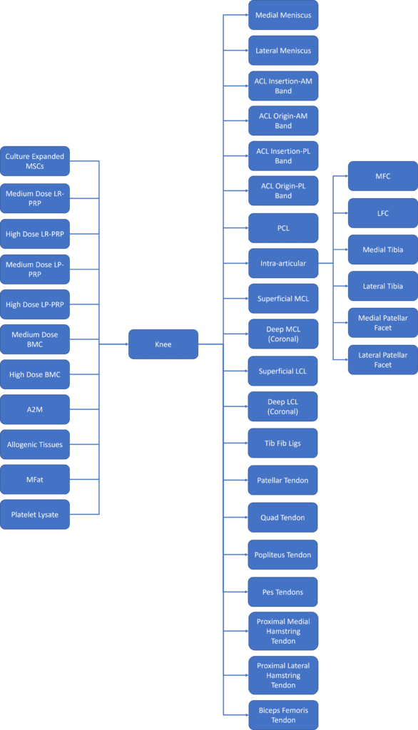
I’m a visual person, so I love diagrams. Check out the one I created below for the knee:

On the left, you see the 11 choices I have as a doctor on which Orthobiologic to use. On the right, you see the 20 places just within the knee where these substances can be placed. Then on the far right, you see just the subchoices inside the joint space and that includes placing Orthobiologics in the bone (Intraosseous). If I did my math right, choosing these sites randomly without understanding how to read your own MRI images would result in a bit under 200,000 combinations. Meaning you would be VERY unlikely to be able to place the right stuff in the right place without understanding history, exam, and imaging.
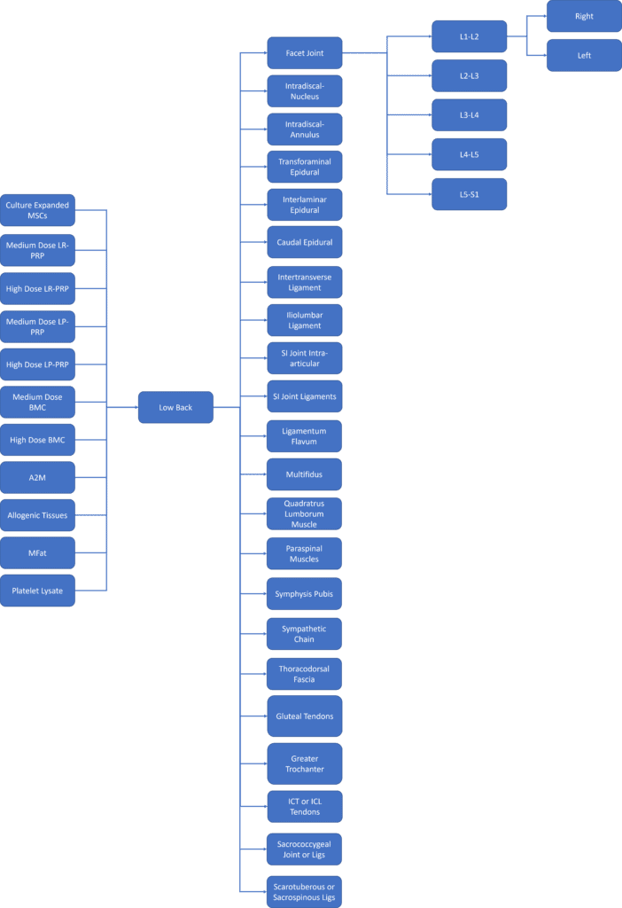
Let’s take the low back:

Same deal. Lots of choices of Orthobiologics and lots of places they could end up. Again, I can’t emphasize enough, being able to know what to place where improves your chances of getting better. A doctor who relies on MRI reports where most of these structures aren’t even mentioned is doomed to be a mediocre provider who is less likely to be able to help you.
How Reviewing MRI IMAGES Improves Therapeutic Accuracy – A Case Report

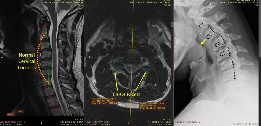
This morning I’ll go over the case of a gentleman who showed up to my clinic with chronic neck and headache pain. He had seen many different providers who never discussed that he had an obvious problem at C3-C4. Why? Because they all relied on the MRI reports!
Looking at the images above, note the one on the left shows the normal curve in the neck as a dashed line. Note that his spine goes backward and that’s centered at C3-C4. So we have our first clue that C3-C4 may be worth our time checking out. The problem, the MRI report never mentioned this curve reversal or the fact that it was centered at that level.
Next, looking at the Axial “top-down” image of each level, we can further focus on C3-C4. In the middle is his Axial MRI image at that level which shows a very large C3-C4 facet joint on the left of the image, That joint is about 3X the size of the one on the right of the image. That means that this Facet joint has significant arthritis. Why? That’s a very strange level to see this as wear and tear arthritis is usually lower down. Maybe there is a movement-based image that shows why that happened?
The Flexion-Extension x-ray on the right tells us why C3-C4 is breaking down. He’s unstable there with the C3 vertebra going forward of the C4. Note the dashed black lines at the back of the vertebrae don’t line up at that level.
All of these imaging findings matched up with his complaints and physical exam. He was tender over this specific arthritic Facet joint and had pain in the area where that joint refers. Hence the diagnosis is chronic instability at C3-C4 causing Facet arthritis at that level and likely irritating the exiting C4 nerve.
What can be done? Obviously, we need to direct our attention to c3-c4. Ablative treatments like Radiofrequency (RFA) will only make his instability worse. While he may need a surgical fusion at that level before that’s needed we would want to try injecting his own platelets (Medium-dose LP PRP) into the ligaments and that arthritic Facet joint (High-dose LP-PRP).
You Should Be Shown What’s On Your MRI By Your Doctor
One of the things I always do during an initial evaluation is to sit down with the patient and read their MRI and other imaging with them. This helps me be able to slow down and actually look at all of the images and it helps the patient understand what’s wrong. This is a critical step as an educated patient is a better patient than one who has no idea what’s wrong or how I plan to help.
Hence, if this doesn’t happen, your first question to your doctor should be, “Did you personally review all of my imaging by reading the actual films?”. If the answer is, “I read the reports” then find a new doctor. If the answer is “Yes”, then ask to be shown in the office or through a telehealth visit what’s on your imaging. Tell your doctor you want to be educated about what’s wrong. If your doctor can’t take the time to do this, then again, find another doctor.
A Puzzle Piece is Not the Whole Puzzle!
While I have extolled the virtues of a doctor who understands that reading imaging studies are key to planning the right Orthobiologics treatment, there is a word of caution required. Remember when I said that imaging was one part of getting to a diagnosis and planning treatment? If the doctor doesn’t also take an extensive history and perform an extensive hands-on exam, then imaging is merely a single puzzle piece. Let me explain.
How many times have you been working on a puzzle when you see one piece you’re convinced fits into the rest of the puzzle, but then it doesn’t really fit that spot? We all know that you can’t tell what image the puzzle shows until most of the pieces have been assembled. In the same way, an overreliance on imaging without a thorough history and hands-on physical exam can lead to the wrong conclusions about diagnosis and treatment. Hence, you want a doctor to read their own images and also spend valuable time with you getting a history and performing an extensive hands-on exam.
The upshot? Imaging is a key part of figuring out what’s wrong and how to treat it. Hence a doctor that reads their own imaging in front of you is a valuable asset to have on your side. A doctor who only knows how to read radiology reports is more likely to come to the wrong diagnosis and prescribe the wrong Orthobiologic treatment!

If you have questions or comments about this blog post, please email us at [email protected]
NOTE: This blog post provides general information to help the reader better understand regenerative medicine, musculoskeletal health, and related subjects. All content provided in this blog, website, or any linked materials, including text, graphics, images, patient profiles, outcomes, and information, are not intended and should not be considered or used as a substitute for medical advice, diagnosis, or treatment. Please always consult with a professional and certified healthcare provider to discuss if a treatment is right for you.
