What Are the Intertransverse Ligaments?
As I have said many times, ligaments are the red-headed stepchild of the spine. Meaning they are often ignored by 99% of the physicians treating spine problems. However, they are important and ubiquitous throughout the spine. Today we’ll review the Intertransverse ligaments, what they do, and why they could be causing you problems. Let’s dig in.
The Spine Ligaments
The number of ligaments in the spine is shocking. Just take a look at the image I prepared below:
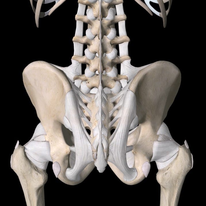
This is the Pelvis and Lumbar spine. Look at the ligaments around the hip joints, SI joints, and lowest Lumbar levels. Like a building where there is more need for heavy structural support at the bottom, the Lumbar spine is dense with ligaments that form that base of support. As you go up the spine, just like in a tall building, while the amount of ligament structural support gets less, it’s no less important. Take a 100 story skyscraper. If you remove some of the structural steel on the 60th floor, you’re going to have serious problems. Hence, in the spine, if you damage the ligaments in the Thoracic spine (upper back) or Cervical spine (neck), you will have big problems supporting the remaining structure.
The Intertransverse Ligaments
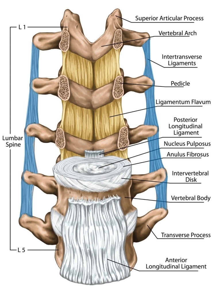
Staying with our skyscraper analogy, let’s look at what kinds of support are needed. In our tall building, you might imagine that you need central structural support to tie into. In a building, that’s the central core that contains the elevators and stairs and that’s constructed of steel-reinforced concrete. In your body, the spinal bones serve that purpose. The difference is that in the skyscraper, nothing is supposed to move very much (other than some small amount of sway in the wind). In the spine, the whole central core structure is built to twist and bend, which makes the supports all the more complex.
In the building, the steel that supports any floor comes off of the central concrete core. In the spine, you also have ligaments that serve that same purpose. One of those that provide lateral side-to-side stability is called the Inter-transverse ligaments.
In the diagram I have prepared above, note that these Intertransverse ligaments are highlighted in blue. They go from vertebra to vertebra connecting the bony projections on the sides called transverse processes. These ligaments help to primarily provide resistance to side-to-side bending and secondarily the rotation of one vertebra on the other.
Intertranverse Ligament Problems
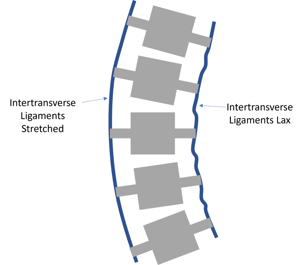
Perhaps the most visible issue with Intertransverse ligaments is Scoliosis or side bending and rotation of the spine. As shown above, in this disease process, the Intertransverse ligaments on the side of the convexity are stretched. On the side of the concavity, they’re lax. This is one of the things that allows side bending to happen. In that case, to treat Scoliosis through injections, we inject those stretched ligaments in a procedure called a percutaneous scolioplasty.
Another thing that can happen is that the Intertransverse ligaments can be injured in a traumatic event. That can lead to Scoliosis or just lateral instability that’s worse with side bending.
Injecting the Intertransvse Ligaments
Injection of the Intertransverse ligaments can be accomplished using x-ray (fluoroscopy) or ultrasound guidance. For patients who are thinner, they can be seen on ultrasound. For heavier patients or in the Cervical spine (where the vertebral artery is close), fluoroscopy is needed. They can be easy to inject or in areas like the neck, time-consuming and difficult because of the critical artery nearby. If fluoroscopy is used, x-ray contrast injectate must also be used to make sure that the doctor is in the right spot. This is important as many prolotherapists that use fluoroscopy don’t inject radiographic contrast to make sure they’re in the ligament and not in the vertebral artery.
Injection choices span the gamut of any ligament injection in the spine. Basic prolotherapy may work but require more sessions. PRP can be helpful as well and usually takes fewer injection sessions. Bone Marrow Concentrate is also an option in patients with recalcitrant problems.
Intertransverse Ligaments at the Top
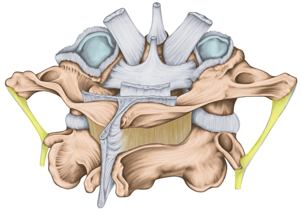
The Intertransverse ligaments run all the way up and down the spine. At the very top of the neck, they become important as shown above as the body has a bowling ball on the end of a stick problem. Meaning the head is heavy and supporting it takes many ligaments as shown above. Shown are the C1 and C2 bones with the head removed. Note all of the ligaments here (Alar, Transverse, Apical, PAOM). Also, note the Intertransverse ligament I have highlighted in yellow. Take a look at how that now flares out further to the side, to begin to prepare to deal with the bigger loads that the head will create.
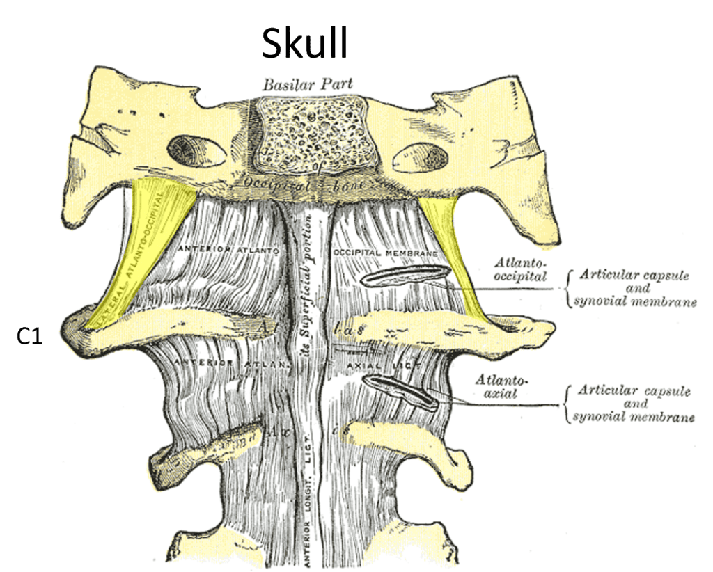
When the Intertransverse ligament hits the very top, it’s called the lateral Atlantooccipital ligament and connects C1 to the skull. I’ve seen a reasonable number of Craniocervical Instability patients have injuries here. This injection is VERY complex as the vertebral artery is very close here, so it requires not only C-arm fluoroscopy to guide the needle and contrast injection to make sure that the injection isn’t into the artery but the intermittent use of a technology known as Digital Subtraction Angiography (DSA). This allows the physician to see subtle take up of radiographic contrast and avoid a HUGE problem. Hence, this ligament should never be injected using ultrasound only or just fluoroscopy without contrast and DSA.
The Intertransverse Ligament at the Bottom
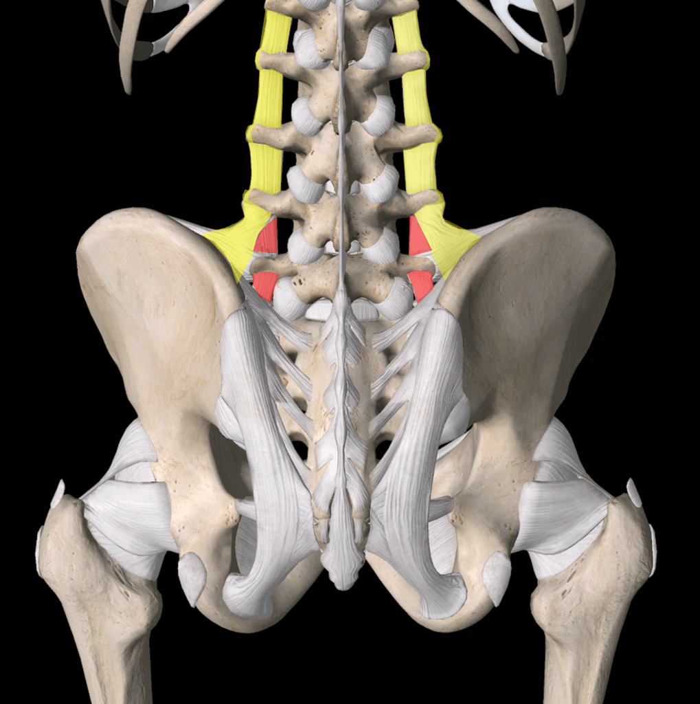
Here I’ve highlighted the Intertransverse ligament down to the bottom of the spine where it meets the Pelvis. The yellow goes down to become the Iliolumbar ligament which attaches to the Pelvis and actually connects into the front part of the Sacroiliac joint. The red here at the bottom represents the same ligament direction that the Intertransverse takes in the rest of the Lumbar spine. Hence, at the bottom, it also flares out and bifurcates into named ligaments. However, these ligaments are all confluent with each other.
A patient with an Intertransverse or Iliolumbar ligament issue here will have problems with sideways bending and may have been in a crash where the impact came from the side. These ligaments also help to stabilize the L5 in forward motion, so in patients with a slipped L5 (Spondylolisthesis), that can be due to a lax Intertransverse or Iliolumbar ligament. Again, these ligaments can be injected as above.
The upshot? The Intertransverse ligament is often ignored, but it should be one of the things that you and your doctor consider in your journey from chronic spine pain to getting better!

If you have questions or comments about this blog post, please email us at [email protected]
NOTE: This blog post provides general information to help the reader better understand regenerative medicine, musculoskeletal health, and related subjects. All content provided in this blog, website, or any linked materials, including text, graphics, images, patient profiles, outcomes, and information, are not intended and should not be considered or used as a substitute for medical advice, diagnosis, or treatment. Please always consult with a professional and certified healthcare provider to discuss if a treatment is right for you.
