How to Interpret Cell Viability Studies Used by Amnio and Cord Vendors: Corecyte Review

I would love to find an amniotic, placental, or cord product available on the market today that has millions of stem cells per vial. However, I know too much about this subject to be hoodwinked by orthopedic sales reps trying to move dead cells as a live stem cell product. This morning I’d like to share with you a report sent to me by two physicians this week as evidence that a cord “stem cell” product (Corecyte made by Predictive Biotech) has millions of viable stem cells to teach you how to separate hyperbole from useful data.
What Are Amniotic and Cord Stem Cells?
Every baby delivered has lots of tissues left behind. These include the placenta, the birth sac, amniotic fluid, and the umbilical cord. These tissues can be chopped up, pulverized, frozen and/or bottled. For many years they were specialty tissue products that didn’t go through full FDA approval, but merely a quick registration. Then, a few years ago, a smart sales rep figured out how to take this slow-moving tissue market and turn it into a sales hit. Simply tell doctors there are viable stem cells in this stuff. Regrettably, our tests and tests performed by another organization haven’t been able to confirm that any of these products have live stem cells. However, that hasn’t stopped disreputable companies, sales reps, and doctors from stretching the truth and telling patients that these tissues have live stem cells that can cure what ails them.
The companies vending this tissue usually work through independent orthopedic sales reps whose knowledge of cell-based medicine is scantly more than that of a high-school-biology student. They often tell physicians that these tissues have live stem cells and many put that in writing. This week, two sales reps working for the same company sent reports to two of my colleagues purporting to show that their product has millions of viable stem cells.
Yesterday’s E-Mails and Viability Testing
The two e-mails I received yesterday were from a university-based orthopedic surgeon and a pain-management physician (from two different academic institutions). The product they want me to review is an umbilical cord tissue that we had on our docket to test but had yet to receive a sample from the company (Corecyte made by Predictive Biotech). Most interesting was the fact that these e-mails had attachments that, according to the sales rep, supported that the product had large numbers of viable mesenchymal stem cells. Digging into these reports will help everyone evaluating these products understand how to separate cell therapy facts from fiction.
The University Viability Report
First, companies often use universities to try and bolster their claims. This is because the first thing many physicians will think is, Wow, some smart people at a university tested this and they said it’s great, so it must be great! So it wasn’t surprising to see this newest cord cell product using reports produced by two different universities. However, we’re going to break down these reports based on what they say and don’t say, as I’ve never been one to be impressed by a label.
The first report sent to my colleagues is from a company memo that details simple live/dead staining that the firm had performed on one of its cord tissue products at Ohio State. They ran three tests on samples containing 500,000, 2M, and 5M total cells. The viability ranged from 64–78%. That sounds pretty good, maybe, right? Not so fast.
First, realize that these are total cell numbers and not stem cell numbers. Even in an umbilical cord, the number of stem cells present in any tissue is only a small fraction of the total cells. So we have no idea based on this report how many of these cells are actually stem cells.
Second, to understand the viability numbers reported, I’ll need to use a patient example. A live/dead stain only says that the cell is alive (and sometimes that’s not 100% correct). To help doctors (and patients) understand what these viability numbers mean, I like to use the example of patients who are in various states of health in a hospital.
Using patients as a proxy for cells, the guy in the ICU who is one day away from death’s door is counted as alive—so is the patient in hospice, who is NPO and DNR, as well as a young and healthy kid in the ER with a fracture. However, only one of them will ever have a functional life. The same holds true with cells. So what this live/dead test doesn’t say is as important as what it does. It doesn’t tell us if these cells that are technically alive can actually function. This is why when testing these products, we usually add apoptotic markers that can separate the two almost-dead patients from the vital one and culture expansion to make sure the cells are capable of growing (i.e., leaving the hospital and going back to work).
So can we conclude anything from this OSU testing? One thing I can say is that from 12 years of working with culture-expanded cells that are placed into and recovered from cryopreservation, these viability numbers are low. For example, for autologous cells thawing out of the freezer, you would expect viability to be in the low 90s. Sometimes you’ll see older cells that have a viability in the high 80s. However, if viability runs in the 60s to 70s, then as a physician using cryopreserved cells, I would be very concerned about cell health. So we do have a hint from the cord-tissue testing that these cells have been through the proverbial wringer.
The Second University Report on Flow-Cytometry Studies
If you really want to impress a physician, throw flow-cytometry data at them. This is not something that 99% of doctors get exposed to, so it looks very scientific and usually sails over their head. So it’s not surprising to see flow data reported by the University of Utah in the information sent yesterday to physicians. On the one hand, If it’s run with the right markers and interpreted correctly, it could be very helpful, but if it’s not, that’s a problem.
This second report says that the university tested two samples of umbilical cord tissue provided by the company. One was “fresh” and the other “frozen.” There are two parts to this report: one that has to do with viability using flow-cytometry markers and the other tried to ID how many mesenchymal stem cells are in the sample.
First, the viability testing performed for the frozen sample (which is what the company sells), was dismal. It showed viability of 43.1% (versus 67.6% for “fresh”). Even the viability of the fresh sample is really poor, so I imagine this wasn’t a sample run down from the OB ward to the lab, but a sample that had been sitting for a while or shipped refrigerated. However, a viability of 43% for the frozen sample shows us that these cells have indeed endured their own special version of hell. When viability is this low, you have to very concerned about whether anything in this sample is, in fact, functional.
The second part of the report is using flow cytometry to ID how many mesenchymal stem cells are in the sample. To understand how this is done, you have to know a bit about flow cytometry. This technology uses fluorescent stains that bind to specific cell-surface markers. These cells are then lined up one by one and run through a laser to excite the fluorescent dye on an antibody, and a detector counts the hits for each marker. The problem for MSCs is that there is no one marker for this cell type. Hence, the ISCT has produced a position paper that in order to ID an MSC you need the following: CD105 CD45 CD73 CD34 CD90 CD14 or CD11b. Each of these cell-surface markers is also found on lots of other cell types, hence, the reason you need a collection of markers. The CD105, 90, and 73 markers have to be present, and the CD34, 45, and 14 markers have to be absent.
The University of Utah report has issues. First, they only used four markers—CD105, 90, 73, and 45. Second, a glaring error is that they didn’t use the exclusion of CD34 (remember, it has to be absent). This is an issue because that marker is found on hematopoietic stem cells (HSCs), which would be expected to be very numerous in an umbilical cord sample. So without using at least three positive markers and at least three negative markers (i.e., the absence of those markers), there is no way to tell how many MSCs are in this sample.
Despite this issue, the report says there are 800,000 MSCs per ml! This is strange as the report also says that there are only about 800,000 total cells per ml in the sample. So the report says that 100% of the cells are MSCs? That’s bizarre as the MSCs would need to express at least three of the positive markers simultaneously that were represented on only 28–84% of the cells tested. Hence, if we only use positive markers, the least reported marker would be the max MSC percentage, which is 28%. However, since the negative marker for CD34 wasn’t reported (and the CD45 marker isn’t reported as a negative marker in this report), it’s likely that a good number of the cells identified as MSCs were really HSCs and/or more-common white blood cells. So how many viable MSCs were in this sample?
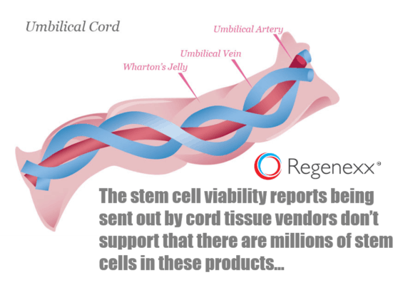
Let’s start with how many MSCs could be in this sample if it included Wharton’s jelly (the largest repository of MSCs in the umbilical cord). This reference discussing the MSC content gives us an idea: “The isolation efficiency from Wharton’s jelly…ranges from 1 to 5 × 10⁴ cells/cm of umbilical cord.” The average cord is 55 cm long, and given the expense of donor testing per cord for communicable diseases, it’s unlikely that more than one cord was used in this product. This means that if we take the average number above of 25,000 per cm X 55cm, we get 1.37 million. However, using centrifugation and washing steps, it would be very difficult, if not impossible, to isolate all of the cellular material in 55 ccs of Wharton’s jelly and cram that into a 1 ml sample. If the company did that, given an MSC percentage of even 10%, we would expect to see some 13.7 million cells in this sample. That wasn’t the case, given that the frozen sample only had about 1.7 million total cells.
However, the company has a stem-cell-isolation problem on its hands because the FDA only allows it to use simple steps, like washing, centrifugation, or adding water. The MSCs from the richest site of the umbilical cord (Wharton’s jelly) can only be isolated using either enzymatic digestion or an explant culture, both of which are off limits. Also, using centrifugation and washing steps, it would be tough, if not impossible, to isolate all of the cellular material in 55 ccs of Wharton’s jelly and cram that into a 1 ml sample. If the company did that, given an MSC percentage of even 10% (extremely high), we would expect to see some 13.7 million cells in this sample. That wasn’t the case, given that the frozen sample only had about 1.7 million total cells.
If we use the 800,000 viable cells/ml number given by the report as representing the total cells in this tested sample, we can get closer to the max number of MSCs that could have been present. At most, the MSC number could have been 28% of 800,000, or 224,000. Again, as discussed, given that the Univerity of Utah didn’t run the appropriate panel to identify MSCs, this lower number likely includes MSCs, some white blood cells, and HSCs.
What Needed to Be Tested but Wasn’t…
So we know that the viability of these cells is poor, and we know that this likely represents a serious challenge to their health, as properly stored and recovered autologous MSCs from older patients have viability rates of >85%. So the next step in testing (not done here) is to see if these cells can still function. At a minimum, we need to determine if they can adhere in culture and replicate under ideal conditions (which they likely won’t encounter in the body once injected). You could also look at the ability to suppress inflammation and differentiate, but suffice it to say that we’ll take even the most basic test that confirms that the cells are functional.
So was this done? No. There are no culture-based tests in either report.
My Conclusions on the University Tests
The sales rep for Corecyte that sent an e-mail to one of my colleagues wrote the following:
“I have just received the enclosed reports which is what you requested of me and our new stem cell injection product from xxx.
XXX has just published two independent lab sources on MSC viability of YYY.
One is from University of Utah and another from Ohio State.
These seem to confirm a range of 800k to 1.2mm MSC’s per vial of YYY.”
So is this backed up by the reports from the two universities? Nope. As discussed, only simple live/dead data was reported, and there was no information to support that there were 800,000 to 1.2 million viable MSCs per vial. In fact, the 1.2 million number seems to have been taken from the fresh cell sample tested by the University of Utah! Given that this company doesn’t sell a fresh version of this product (only frozen), it seems the sales rep had no idea what he was quoting.
The upshot? While I’m open to any company letting us test their cord-tissue product to try to confirm that these products contain even 500,000 MSCs per vial, this data circulated by this company is disappointing. The good news for the company is that most physicians have no idea what they’re looking at; hence, it’s easy to wow them with reports from a university lab. However, if you’ve read this blog, now you know how to question sales reps and the reports they send! In the meantime, we’d still love to test some of this cord product or any other product. We’re still hoping that someone out there has a product that actually has millions of stem cells.

NOTE: This blog post provides general information to help the reader better understand regenerative medicine, musculoskeletal health, and related subjects. All content provided in this blog, website, or any linked materials, including text, graphics, images, patient profiles, outcomes, and information, are not intended and should not be considered or used as a substitute for medical advice, diagnosis, or treatment. Please always consult with a professional and certified healthcare provider to discuss if a treatment is right for you.
