ACL Surgery Alternative: Imaging Analysis in our New Stem Cell ACL Tear Studies
Can a stem cell injection be an ACL surgery alternative? The ACL is the major stabilizing ligament of the knee. ACL tears are a big deal, especially in young athletes. In women’s soccer, torn knee ACLs seem to be an outright national epidemic. The surgery for this procedure is very invasive and installs a new ligament without many of the natural properties of the original equipment. Healing an ACL tear would be nothing short of miraculous, allowing the athlete to keep that ligament. But can this be done? We’ve been seeing evidence of healing of torn ACLs for years using a very exacting injection of stem cells directly into the torn ligament. To prove the point, we’re now funding two studies to make sure that the data gets into the peer reviewed medical literature.
When a doctor looks at an MRI of the ACL, it’s a very qualitative thing, meaning whether the ACL is healed is based on how the picture looks and how a reader interprets that image. However, research journals like quantitative data like blood tests and other diagnostics that spit out a number. So is there a way to bridge that gap between the two and use a quantitative way to see if the ACL ligament is better after a stem cell injection? Yes. Our research team is using MRI analysis software to turn the ACL before and after images into pixel intensity histograms.
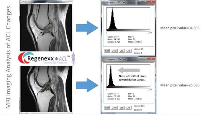
A histogram is a graph of the frequency of the appearance of something in a data set. Since we know that on the images where we see the ACL a better ligament is darker and a weaker or damaged ligament is lighter, the imaging analysis software plots the number of pixels in the ACL based on how light or dark they are. A shift toward dark pixels (for our purposes the left skew of the data) tells us in a quantitative way that the ACL looks better.
Look at the images above (click on the thumbnail to see a bigger PDF). Note that the histogram graph for the image has the darkness of the pixels on the x-axis and the number of pixels that were that shade of black, grey, or white on the y-axis. So the position and height of each of these bars is important. Now look at mean of the above histogram, the average pixel position is about 34 1/2. Now look at the histogram below that was created from an image taken a few months after the stem cell injection. The mean has shifted to the left (darker pixels) and is now a bit more than 25. This means that the average pixel value in the ACL got darker from the before image to the after image.
We’ve used this new imaging analysis technique on a number of patients and are readying a case series with objective measurements in ACL stem cell treated knees for submission to a medical journal. Since we’ve validated this method and it has very high inter-rater reliability (Correlation Coefficient >0.9), we’ll use it again for our knee ACL tear randomized controlled trial that’s now recruiting. The upshot? We’re seeing torn ACLs look much better on MRI with very precise stem cell injections and these patients are also reporting improvement. Again, our goal is nothing less than changing how Orthopedic care is delivered, so substituting a specialized injection for a big knee surgery is a good start!

NOTE: This blog post provides general information to help the reader better understand regenerative medicine, musculoskeletal health, and related subjects. All content provided in this blog, website, or any linked materials, including text, graphics, images, patient profiles, outcomes, and information, are not intended and should not be considered or used as a substitute for medical advice, diagnosis, or treatment. Please always consult with a professional and certified healthcare provider to discuss if a treatment is right for you.

