Culture Expanded Stem Cells for Knee Arthritis

©Regenexx
I evaluated an interesting patient down at our Cayman Islands advanced stem cell practice site recently in that his MRI changes were fascinating. While I see patients every day who are improving and have great stories, many times I just don’t have the time to write up what’s happening. However, every once in a while, I see something that’s really interesting and want to share. Since this patient’s MRI shows some amazing post-injection improvements, it’s worth sharing this morning. In particular his knee MRI shows improvements in his knee meniscus and bone.
Why Arthritis May Be More About the Bone than the Cartilage
Simple messaging works in medicine, even though it may not be accurate. Take, for example, the idea that high cholesterol is bad and that you need drugs to lower it. The messaging is simple, but now inaccurate. Turns out that for most of us, high total cholesterol isn’t associated with heart disease, and drugs to lower it have barely there effects. However, that doesn’t stop Madison Avenue from pushing that powerful messaging.
The same holds true for knee arthritis. The simple message is that cartilage cushions the joint and that when it wears out, the joint hurts. However, regrettably, that’s not necessarily true. For example, if you were to take MRIs of the knees of 100 middle-aged or elderly people and only half had knee pain, you would struggle to place the patients with knee pain in the right category just by looking at the status of their cartilage loss on the MRIs. Why? Turns out that cartilage loss is a pretty poor predictor of who has knee pain. Why then do doctors seem to have a cartilage-loss fetish? Like many things in medicine, our focus is misguided by the business of cartilage repair.
If you can’t predict who has knee pain just by looking at the knee MRI image and looking for lost cartilage, is there something else that can predict knee pain more reliably? Yes, it’s called a bone marrow lesion (BML, or also known as bone marrow edema, or BME). This is a swollen area in the bone where there are likely small microfractures occurring. It used to be thought that this happened because of lost cartilage, but we now know that this occurs because of a complex interaction between the bone and cartilage. We also know that these areas hurt.
In conclusion, if you had to look at one thing on a post stem-cell-injection MRI that was really exciting, getting rid of these painful BMLs would be it. This is exactly what happened on this patient’s MRI. However, before we get into what changed, you need to learn a little more about the knee meniscus.
Chopping Up the Meniscus?
This patient had a meniscectomy, meaning that a part of his meniscus was removed because it was torn. Despite the fact that multiple high-level research studies have shown this to be a useless and ineffective surgery, it’s still being performed worldwide. The meniscus is half a ring of spacer tissue, so one of the things that happens when you cut out pieces of it is that the ring weakens. This results in parts of this ring-shaped structure extruding outside the joint, which then causes more pressure on the cartilage surfaces. This last change in the function of the meniscus is well documented in research studies showing a dramatic rise in contact pressures of the joint as more and more meniscus is removed.
Interesting MRIs
This patient is an active middle-aged guy who noted marked problems running and exercising after knee meniscectomy surgeries. He was noted to have arthritis on the inside of his knee joint and a meniscus that was extruding outside the joint and leaving it unprotected. His first treatment was with culture-expanded stem cells at the Grand Cayman advanced-practice site, and his opposite knee responded with about 80% improvement. However, while that knee had a meniscus that was minimally extruded, the side that didn’t respond had an even bigger meniscus extrusion issue. That knee also had a BML, so Dr. Schultz treated both the bone and the meniscus at a second treatment session by precisely injecting culture-expanded stem cells into these areas. This included placing a needle under fluoroscopic guidance into the damaged bone. He was back for another treatment on that knee when I saw him this last week, as he’s been getting incremental improvement with each round of injections.
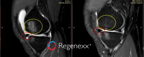
Note the before and after MRIs below, which show his BML. On the left, note that the bright spot in the dark bone that’s in the yellow-dashed line changes from the before to after image. This area becomes normally dark, meaning no more BML. There is now a BML on the tibia, which will be treated this trip.
Also note that there is another curious change. The meniscus position in the before image on the left (the dark triangle-shaped structure in the red-dashed circle, above) is out of the joint and displaced. In the after image, the meniscus is now tucked tightly inside the joint where it belongs. This is likely due to the fact that Dr. Schultz precisely injected the meniscus as well, with the goal of getting better quality and tighter tissue.
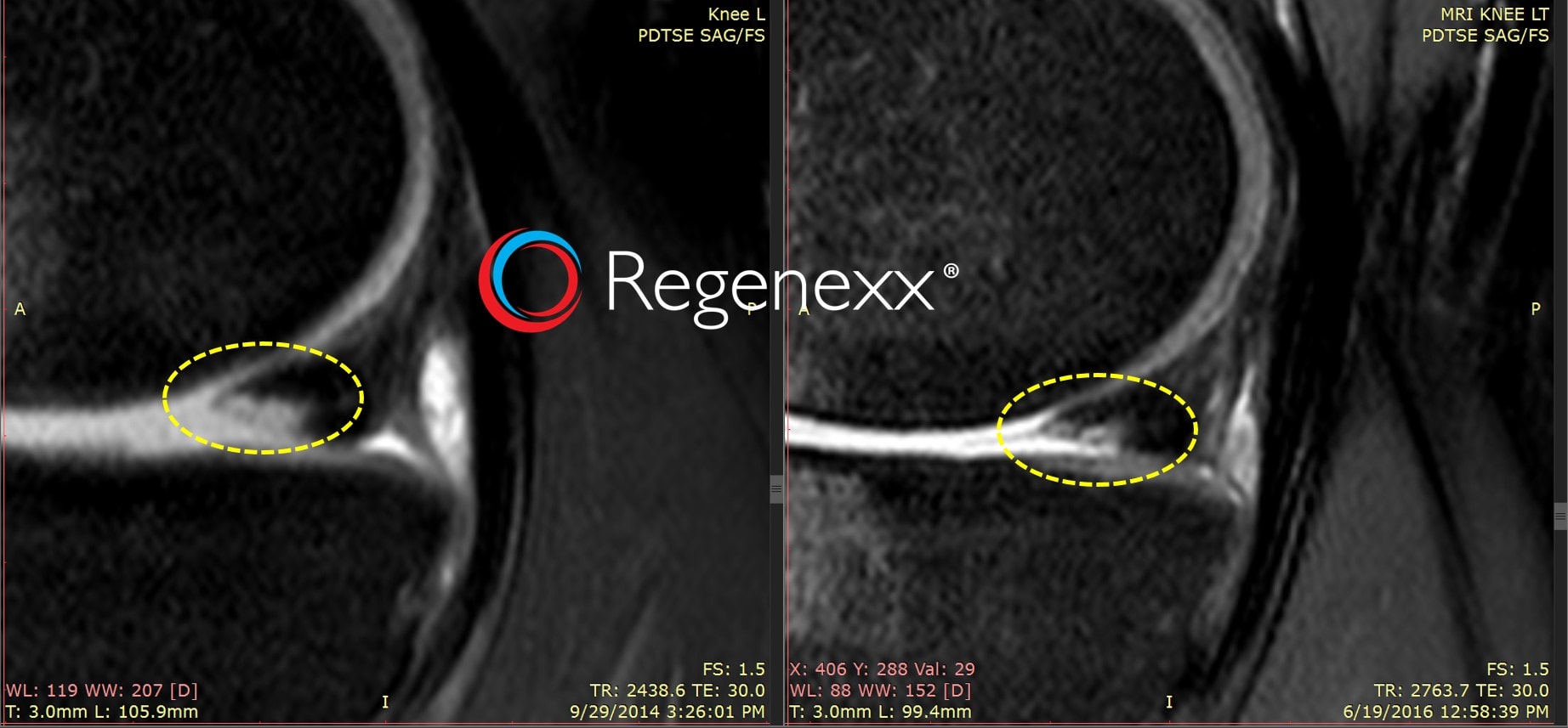
There have also been positive changes in the posterior meniscus MRI as well. Note the large bright spot in the dark triangle in the left before image (yellow-dashed circle). Note that this is smaller in the after image on the right. This is consistent with meniscus repair because normal meniscus signal on MRI is dark. Both of these improvements on imaging are consistent with his reported clinical improvement.
The upshot? While not all patients who get relief see changes on their MRIs, it’s always interesting to see what changes and why. In particular, improvement in painful BMLs are a consistent finding. The meniscus repositioning is also something we’ve been paying closer attention to this past year. We hope that this patient can continue to get great improvements!
The Regenexx-C procedure is not approved by the US FDA and is only offered in countries via license where culture-expanded autologous cells are permitted via local regulations.

NOTE: This blog post provides general information to help the reader better understand regenerative medicine, musculoskeletal health, and related subjects. All content provided in this blog, website, or any linked materials, including text, graphics, images, patient profiles, outcomes, and information, are not intended and should not be considered or used as a substitute for medical advice, diagnosis, or treatment. Please always consult with a professional and certified healthcare provider to discuss if a treatment is right for you.

