What Is a Flexion-Extension X-Ray?
On this page:
- What is spinal instability?
- Can instability be seen on an x-ray or MRI?
- What is a flexion-extension x-ray?
- The problem with flexion-extension x-rays
- How to make sure your test is worthwhile
Back and neck pain patients and doctors are often confused by the idea of instability in the spine. From the patient’s standpoint, since they’ve mostly heard about herniated discs and compressed nerves, this idea is a bit foreign. Doctors are primarily looking for things that show up on MRIs, so they frequently miss the concept of spine instability altogether.
Despite this, finding out if your back or neck is unstable is often critical for finding relief. The test that can discover instability is called a flexion-extension x-ray and despite this being common, it’s usually performed so badly that the results are worthless. Here’s how you can make sure that your test is done right.
What Is Spinal Instability?
Your spine is made up of the neck (cervical), upper back (thoracic), and low back (lumbar) areas. One of the biggest challenges of walking upright on two legs, rather than on four, is keeping the spine stable. Think of it this way–your spine is made up of 24 vertebrae that are stacked like kid’s blocks. When was the last time you stacked more than 20 blocks on top of each other without the whole rickety tower collapsing?
To keep the spine from collapsing, you have two main systems. One is strong ligaments that limit motion in certain areas, sort of like flexible duct tape between the blocks. The other is made up of stability muscles (called multifidus) which help adjust one vertebrae on the other as you move.
If the ligaments get injured or the muscles go off line, the vertebrae can move too much, leading to too much motion between them, or an unstable spine (1). This is a big deal, as unstable vertebrae can cause pain by placing excessive wear and tear on the spine joints (facets) or disc, and can irritate or pinch the spinal nerves leading to nerve pain or sciatica. In fact, sometimes the spine looks fine on MRI when the real culprit is instability.
Can Instability Be Seen on an X-Ray or MRI?
Since regular x-rays and MRIs are static images without any movement, these tests can’t identify instability, which happens only with movement (2). Hence, many patients who have normal or unimpressive x-rays or MRIs are later diagnosed with instability as the cause of their back or neck pain.
What Is a Flexion-Extension X-Ray?
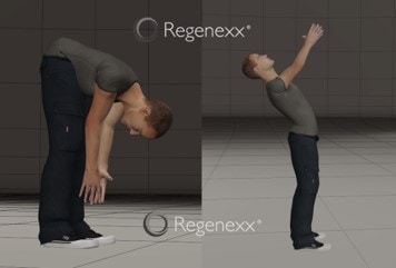
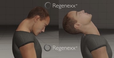
In order to replicate the conditions under which there is too much movement in the spine vertebrae, an x-ray can be taken when the patient moves. This is called a flexion-extension x-ray. For the low back, the patient is asked to bend forward and then backwards while x-ray images are taken in both positions. For the neck, the patient looks down and then up.
The Problem With Flexion-Extension X-Rays
In a perfect world, the technologists taking these images would perform each one with enough movement to show if any instability is present. All too frequently, this doesn’t happen. In fact, most of the time they do it wrong. Why?
Radiology techs get in trouble if a patient reports that this test flared up their pain. Hence, they learn over time that pushing patients to their limit on this test is not advisable. In addition, having the patient get the needed motion may be more technically difficult for the tech. However, if there’s not enough motion, the test can be a “false negative.” This means that instability may be present, but the test will miss it.
How to Make Sure Your Test Is Worthwhile

So for a low back test, for the first
The upshot? A simple test may be able to help your doctors figure out if you have instability causing your neck or back pain. However, knowing how to make the test work to get real results is critical to figuring out what’s wrong!
__________________________________________________
References
(1) Offiah CE, Day E. The craniocervical junction: embryology, anatomy, biomechanics and imaging in blunt trauma. Insights Imaging. 2017;8(1):29–47. doi:10.1007/s13244-016-0530-5
(2) Silvis ML, Mosher TJ, Smetana BS, et al. High prevalence of pelvic and hip magnetic resonance imaging findings in asymptomatic collegiate and professional hockey players. Am J Sports Med. 2011;39(4):715-721. doi:10.1177/0363546510388931

NOTE: This blog post provides general information to help the reader better understand regenerative medicine, musculoskeletal health, and related subjects. All content provided in this blog, website, or any linked materials, including text, graphics, images, patient profiles, outcomes, and information, are not intended and should not be considered or used as a substitute for medical advice, diagnosis, or treatment. Please always consult with a professional and certified healthcare provider to discuss if a treatment is right for you.
