How Good are Hip X-rays and MRIs In Diagnosing the Cause of Hip Pain?
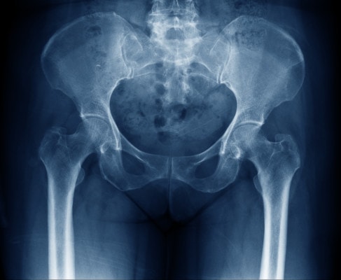
Imaging by itself is often useless in determining why someone has pain. Despite that fact, patients often believe the opposite, that their x-ray or MRI study is a critical tool for making the diagnosis of why their knee or hip hurts. Today we’ll explore research showing that hip x-rays and MRIs are often awful at diagnosing the cause of hip pain. Let’s dig in.

Credit: Shutterstock
Pain and Imaging 101
We’ve known for a long time that images like x-rays and MRIs are really bad at predicting who has pain and who doesn’t. Despite this, many invasive surgeries are launched solely based on an x-ray or MRI result and a cursory 2-minute exam in the office. Let’s explore this concept.
One of the biggest medical scams of the twentieth century that’s still ongoing is a knee MRI that shows a meniscus tear. This one usually begins with a middle-aged patient who feels sudden knee pain when exercising. An MRI shows a meniscus tear and after a cursory exam, surgery is recommended. A part of the meniscus is then removed with an arthroscopic procedure, but the knee pain often persists.
Why is this a scam? Because the research on degenerative meniscus tears is crystal clear and has been for some time (1,2). They are about as important as the wrinkles on your face. Meaning they are ubiquitous in middle-aged and older people who don’t have knee pain. Hence, the fact that a meniscus tear was seen after the knee began to hurt is a red herring and usually not the cause of the knee pain. We also know through multiple randomized controlled trials that any surgery to remove the torn part of the meniscus doesn’t work better than physical therapy or a placebo procedure! (3-7) Finally, we know that this surgery (partial meniscectomy) increases the likelihood of the patient developing knee arthritis (8). Hence, one bad association between a meniscus tear seen on MRI that has been there for years and sudden knee pain leads to a cascading series of bad decisions that cause patient harm.
The Research on Hip Pain and X-rays
Remarkably, despite all that we know about the disconnect between what we see in images and pain, many orthopedic surgeries are still launched based on imaging findings. Take for example the many hip pain patients that I see in the office who have had a hip replacement, based only on an x-ray and cursory exam.
For us physicians to rely on hip x-rays to make big clinical decisions like hip replacement surgery, a few things need to be true. First, people with hip pain must have a high likelihood of having arthritis on their hip x-rays, and conversely, people with no hip pain should mostly have clean hip x-rays. However, what if the opposite was true? That people with hip pain often have clean x-rays and that people with evidence of arthritis on their hip x-rays often have no hip pain?
The research we’ll review is based on data from two government-funded studies: the Framingham Osteoarthritis Study and the Osteoarthritis Initiative Study. You may recognize the name of the first study as it’s the same town in Massachusetts where the famous heart study was conducted. Let’s dig in.
In the Framingham study, the researchers looked at the hip x-rays of 946 patients. Only 16% of hips in patients with frequent hip pain had x-ray evidence of hip arthritis. On the other hand, only 21% of the patients with hip arthritis on x-rays were frequently painful. Let’s let that sink in for a second. There was a VERY POOR correlation between what the researchers found on hip x-rays and hip pain.
Well, maybe this is just one study? Nope. Another even larger government-funded study (OAI) looked at 4,366 hips. They found that only 9% of hips in patients with frequent pain showed x-ray evidence of hip arthritis, and 24% of hips with x-ray evidence of hip arthritis were frequently painful! Meaning that again, there was a VERY POOR correlation between what the researchers found on hip x-rays and hip pain.
The Implications of this Research
This research is clear, relying on an x-ray showing hip arthritis in a patient with hip pain as the main reason to perform major surgery like a hip replacement is NOT evidence-based medicine. In fact, based on the published research, it’s BELOW the standard of care. Hence if you’re a patient with hip pain that has a surgery planned based on an x-ray, then you need to get a second opinion.
Hip MRIs?
Maybe planning hip surgery based on hip MRI results works better? After all, an MRI shows more than an x-ray. In addition, a common practice these days is to get a hip MRI in a younger patient with hip pain, and if that shows evidence of a labral tear or FAI (impingement) then hip arthroscopy surgery for labral repair or reconstruction is often the next step. Let’s explore that concept:
- This US study looked at high-resolution (3T) MRIs and found that labral tears and hip arthritis were NOT associated with hip pain. What was? Specific cartilage lesions on the socket part of the joint (acetabulum) and swelling in the bone (BML or BME) (10).
- An imaging review of patients who were on average 38 years old with no hip pain showed abnormalities including cartilage defects and FAI (impingement) in almost 70%! (11)
- This research showed that labral tears aren’t associated with hip pain, but more sophisticated cartilage mapping (not usually done on MRI) may be able to find painful cartilage lesions (13).
- This study looked at the non-painful side of a routine hip MRI. Labral tears were commonly found in patients with no hip pain on that side (14).
- An interesting study took patients who had been diagnosed as needing hip arthroscopy due to labral tears/FAI based on MRI and performed numbing injections. Patients with labral tears and FAI showed no correlation between the numbing injection and relief, meaning that these findings didn’t cause hip pain (12).
Hence, the idea that hip MRIs can be used to diagnose a labral tear or FAI in a younger patient with hip pain and that finding should be used to offer these patients surgery is not supported by the existing research.
What Could be Causing Hip Pain?
The obvious question here is if x-rays and MRIs are so bad at diagnosing the cause of hip pain, what else could it be? First, it’s critical to note that diagnosis should always go beyond an image plus a 2-minute exam. Meaning that there is no substitute for time with the patient. The history of how the hip pain began and where it’s located is critical. In addition, a good 10-20 minutes with hands on the patient looking at the hip, the low back, and other areas is critical. Hence, if you got an image and a quick cursory exam, then you have not been adequately diagnosed.
In addition, lots of other things cause hip pain. For example, the SI joint is a major cause. Another is irritated low back nerves. Damaged or inflamed tendons around the hip are also a common cause. Hence there are many things that cause hip pain that can be missed by hip imaging.
The upshot? Finding arthritis on a hip x-ray or a labral tear or impingement on a hip MRI does NOT accurately diagnose why someone has hip pain, so basing surgical decisions on this imaging is foolish. As doctors, we need to stop our reliance on imaging and spend much more time figuring out why the patient has hip pain.
_________________________________________________
References:
(1) Englund M, Guermazi A, Gale D, et al. Incidental meniscal findings on knee MRI in middle-aged and elderly persons. N Engl J Med. 2008;359(11):1108–1115.
(2) Risberg MA, Degenerative meniscus tears should be looked upon as wrinkles with age—and should be treated accordingly. British Journal of Sports Medicine 2014;48:741.
(3) Finnish Degenerative Meniscal Lesion Study (FIDELITY) Group. Arthroscopic Partial Meniscectomy versus Sham Surgery for a Degenerative Meniscal Tear. N Engl J Med 2013; 369:2515-2524
(4) Katz JN, Brophy RH, Surgery versus Physical Therapy for a Meniscal Tear and Osteoarthritis. N Engl J Med 2013; 368:1675-1684
(5) Sihvonen R, Englund M, Mechanical Symptoms and Arthroscopic Partial Meniscectomy in Patients With Degenerative Meniscus Tear: A Secondary Analysis of a Randomized Trial. Ann Intern Med. [Epub ahead of print 9 February 2016]164:449–455. doi: 10.7326/M15-0899
(6) Noorduyn JCA, van de Graaf VA, Willigenburg NW, et al. Effect of Physical Therapy vs Arthroscopic Partial Meniscectomy in People With Degenerative Meniscal Tears: Five-Year Follow-up of the ESCAPE Randomized Clinical Trial. JAMA Netw Open. 2022;5(7):e2220394. doi:10.1001/jamanetworkopen.2022.20394
(7) Katz JN, Brophy RH, Chaisson CE, et al. Surgery versus physical therapy for a meniscal tear and osteoarthritis [published correction appears in N Engl J Med. 2013 Aug 15;369(7):683]. N Engl J Med. 2013;368(18):1675–1684. doi:10.1056/NEJMoa1301408
(8) Roemer FW, Kwoh CK, Hannon MJ, et al. Partial meniscectomy is associated with increased risk of incident radiographic osteoarthritis and worsening cartilage damage in the following year. Eur Radiol. 2017;27(1):404–413.
(9) Kim C, Nevitt M C, Niu J, Clancy M M, Lane N E, Link T M et al. Association of hip pain with radiographic evidence of hip osteoarthritis: diagnostic test study BMJ 2015; 351 :h5983 doi:10.1136/bmj.h5983
(10) Kumar D, Wyatt CR, Lee S, Nardo L, Link TM, Majumdar S, Souza RB. Association of cartilage defects, and other MRI findings with pain and function in individuals with mild-moderate radiographic hip osteoarthritis and controls. Osteoarthritis Cartilage. 2013 Nov;21(11):1685-92. doi: 10.1016/j.joca.2013.08.009. Epub 2013 Aug 12. PMID: 23948977; PMCID: PMC3804140.
(11) Register B, Pennock AT, Ho CP, Strickland CD, Lawand A, Philippon MJ. Prevalence of abnormal hip findings in asymptomatic participants: a prospective, blinded study. Am J Sports Med. 2012 Dec;40(12):2720-4. doi: 10.1177/0363546512462124. Epub 2012 Oct 25. PMID: 23104610.
(12) Kivlan BR, Martin RL, Sekiya JK. Response to diagnostic injection in patients with femoroacetabular impingement, labral tears, chondral lesions, and extra-articular pathology. Arthroscopy. 2011 May;27(5):619-27. doi: 10.1016/j.arthro.2010.12.009. PMID: 21663719.
(13) Grace T, Samaan MA, Souza RB, Link TM, Majumdar S, Zhang AL. Correlation of Patient Symptoms With Labral and Articular Cartilage Damage in Femoroacetabular Impingement. Orthop J Sports Med. 2018 Jun 15;6(6):2325967118778785. doi: 10.1177/2325967118778785. PMID: 29977942; PMCID: PMC6024532.
(14) Vahedi H, Aalirezaie A, Azboy I, Daryoush T, Shahi A, Parvizi J. Acetabular Labral Tears Are Common in Asymptomatic Contralateral Hips With Femoroacetabular Impingement. Clin Orthop Relat Res. 2019 May;477(5):974-979. doi: 10.1097/CORR.0000000000000567. PMID: 30444756; PMCID: PMC6494314.

NOTE: This blog post provides general information to help the reader better understand regenerative medicine, musculoskeletal health, and related subjects. All content provided in this blog, website, or any linked materials, including text, graphics, images, patient profiles, outcomes, and information, are not intended and should not be considered or used as a substitute for medical advice, diagnosis, or treatment. Please always consult with a professional and certified healthcare provider to discuss if a treatment is right for you.
