The Multifidus Muscle and Spine Pain – Cause and Effect
The multifidus muscle is the most important stabilizer of your spine, but one that most patients and physicians have never studied. There are now hundreds of research studies confirming that when this little muscle goes south, all heck breaks loose in the neck and back. The multifidis is easy to spot on the average MRI and is now being looked at by NASA as an issue for long-term spaceflight, but to your average pain specialist, surgeon, or radiologist, it’s pretty much invisible.
The role of the multifidis in pain and spine degeneration
Normal Multifidis Function
First, the multifidus is an absolutely critical muscle that stabilizes your spine. To get a sense of what it does, take a look at the video to the right, which shows what happens to your lower back vertebrae (bones) when you walk. The vertebrae are under constant micro-motion feedback from the multifidus so that motion is allowed, but only a small amount in all of the right directions.
Multifidis Problem Causing Pain and Dysfunction
Now take a look at the vertebrae when the multifidus is off-line in the video to the left. The back bones are out of control and there’s too much sloppy motion. All of the extra movement can damage the disc spacer between the bones and the joints between them (facet joints), causing pinching of the spinal nerves.
The Multifidis on MRI
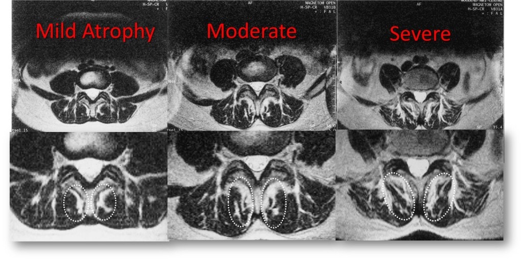
It’s not hard to spot an atrophied low back multifidus on MRI. In fact, it’s easy enough that anyone can do it with just a little training. If you click the thumbnail to the right you’ll see several examples, from a normal multifidus to one that’s severely atrophied. You can also read about how to grade the multifidus on your MRI here. So if you have an MRI of this area, take a few minutes to have another look and see what category you’re in. This alone may be one of the most important things on the film that was never read by the radiologist.
Why don’t radiologists and surgeons pay more attention to the multifidis?
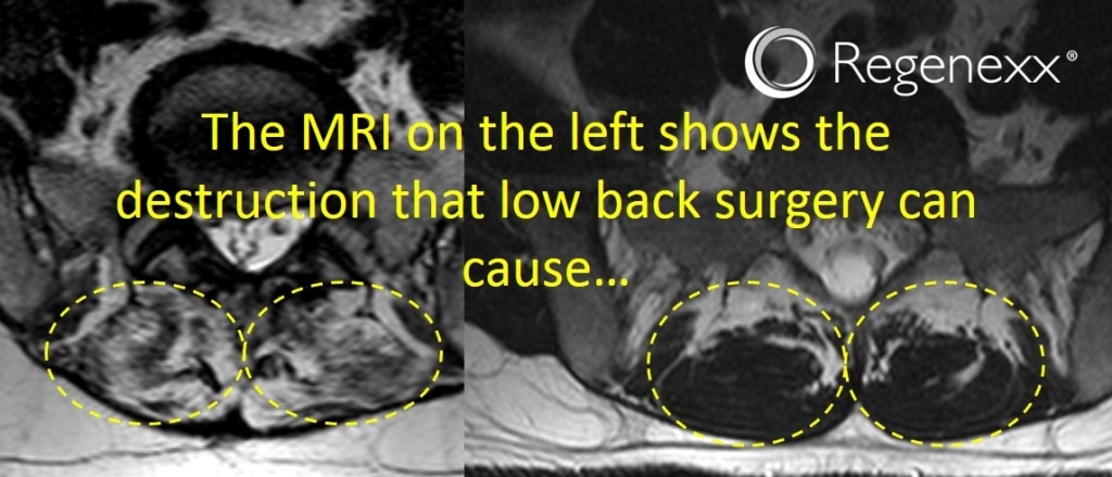
Frankly, it has more to do with the business of medicine than anything else. There really is no surgery for this problem and surgeons are generally locked into a 1970s structural model of the spine, looking for discs and bones pressing on nerves that can be cut out. Radiologists tend to read what surgeons want to see, which is how they garner business. While surgeons may be oblivious to the multifidus, they do a great job of destroying the muscle with their procedures. In fact I just sat and looked at an MRI yesterday of a patient after a low back fusion where multiple levels of multifidus were fried.
While surgeons and radiologists generally ignore this stabilizer, NASA and the European Space Agency are paying attention. A recent study of an astronaut’s multifidus muscle after being on the International Space Station showed that despite special exercises, the muscle did atrophy. Thankfully it returned to normal size once the astronaut was back in earth’s gravity.
More research outside of space continues to be published on the critical muscle, such as a recent study of herniated low back discs. This new study shows that the multifidus goes south shortly after a herniated disc begins placing pressure on the spinal nerve. This makes sense, as the nerve innervates the multifidus, so the muscle shrinks and becomes weaker as it’s nerve supply is disrupted.

NOTE: This blog post provides general information to help the reader better understand regenerative medicine, musculoskeletal health, and related subjects. All content provided in this blog, website, or any linked materials, including text, graphics, images, patient profiles, outcomes, and information, are not intended and should not be considered or used as a substitute for medical advice, diagnosis, or treatment. Please always consult with a professional and certified healthcare provider to discuss if a treatment is right for you.


