Treatments for knee cartilage lesions

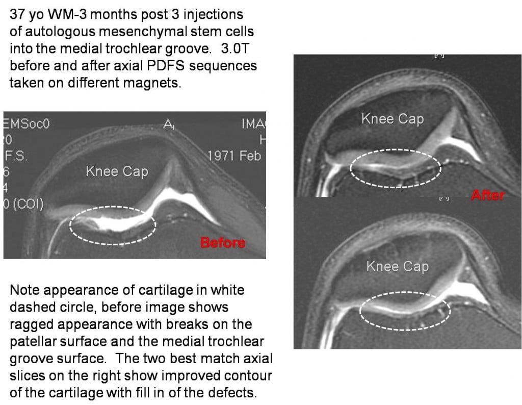
As part of the imaging follow-up series on our current patients, this morning I present the before and after MRI’s of CH. These are axial (saw you in half views) of the knee. The knee cap is labeled. The dashed circle in the before image on the left “BEFORE” images show a ragged appearance of the grey cartilage on the dark bone. The white stuff in between the two is fluid (an effusion or swelling in the joint). You can see the jagged edges of the cartilage and loss of the grey cartilage in the groove (trochear groove). The two after images on the right show a different appearance of this area in the dashed circle. There are two images for the after, as they are a best fit to the same area as the before image (MRI’s “slice” through a certain area, so there are two slices on the right). On the “AFTER” images, there appears to be good “fill in” of the cartilage loss at the trochlear groove. This patient was not treated with micro fracture which would require extensive down time, but this cartilage osteochondral defect was treated by injecting the patent’s own mesenchymal stem cells directly into this lesion on MRI planned fluoroscopy. He had three injections which took place over three months and these “AFTER” images were taken at about 4 months after the first injection.
The knee cap sits in a groove, commonly called the trochlear groove. When there is trauma (such as a knee cap hitting the dashboard) or extensive and excessive wear forces (commonly seen in kicking athletes), the cartilage can wear out such as in CH. We’ve also seen a relationship between patello-femoral syndromes and chronic low back pain. In these cases, the low back impacts the ability to fully contract the quadriceps and ultimately results in pain between the knee cap and it’s groove. Physical therapy can be used to achieve better firing of all parts of the big quadriceps muscle and sometimes this helps. We commonly use IMS to get rid of trigger points in the low back and quadriceps of patients who seem to have a low back problem leading to knee pain. This often makes a huge difference and in our experience, when combined with physical therapy, IMS can usually knock out most patello-femoral syndromes in this category. However, when the problem has been there for many years, a “hole in the cartilage” or OCD (osteochondral defect) can develop. These usually occur in the trochear groove or on the knee cap itself. Surgical treatments can include debridement (cutting out loose pieces), but recent research has shown that arthroscopic debridement is not better than placebo surgery. Other options include micro fracture. This is where the surgeon pokes holes in the bone to try and stimulate healing. This works better in younger patients, but requires extensive down-time on crutches, with about 6-12 weeks of keeping weight off the area. The other option, as shown above, in our opinion, is injecting the patent’s own mesenchymal stem cells into the lesion. With this approach, there is much less down time (we would recommend light walking and aerobic work using an elliptical almost immediately after the procedure).
In summary, the knee cap sits in a groove and can wear down the cartilage in the groove. There are many options for treatment of cartilage lesions in this trochlear groove, including one just involving a series of simple injections.
This patient was treated with the Regenexx-C (cultured stem cell injections). Please note that not all patients with patello-femoral problems can expect to see the same changes on their MRI or the same clinical results.

If you have questions or comments about this blog post, please email us at [email protected]
NOTE: This blog post provides general information to help the reader better understand regenerative medicine, musculoskeletal health, and related subjects. All content provided in this blog, website, or any linked materials, including text, graphics, images, patient profiles, outcomes, and information, are not intended and should not be considered or used as a substitute for medical advice, diagnosis, or treatment. Please always consult with a professional and certified healthcare provider to discuss if a treatment is right for you.
