Alternatives To Knee Replacement Surgery For Osteoarthritis
Knee osteoarthritis (OA) is one of the most common orthopedic conditions. If you are experiencing OA, you understand the pain and discomfort involved and how it can negatively impact your quality of life and enjoyment of daily activities.
Historically, many patients over 40 with knee pain due to osteoarthritis (commonly referred to as arthritis) have been advised to try physical therapy, nonsteroidal anti-inflammatory medications (NSAIDs), hyaluronate or corticosteroid knee injections to reduce pain and inflammation. If these are not helpful, the next recommendation is often arthroscopic knee surgery to repair or remove any damaged cartilage or tissue that may be causing the pain. If the surgery doesn’t deliver the desired results, then total knee joint replacement is usually recommended.
Can Knee Surgery Reduce Pain?
Numerous studies² evaluating the results of common orthopedic knee surgeries have shown that these procedures generally don’t work unless the patient is younger than 40 years of age. Even then, successful outcomes are not guaranteed. If this is new information to you, you are not alone. Many are not aware that surgery isn’t always the solution. Read on to learn more about procedures using Regenexx injectates available at Rehabilitation Medicine Center of New Jersey, NY as an alternative to knee replacement surgery for osteoarthritis.
131 West 35th Street
12th Floor
New York, NY 10001
Request an Appointment
Call to Schedule Schedule OnlineClinic Hours
| Sunday | Closed |
| Monday | 8AM–8PM |
| Tuesday | 8AM–8PM |
| Wednesday | 8AM–8PM |
| Thursday | 8AM–8PM |
| Friday | Closed |
| Saturday | Closed |
How Does The Regenexx Approach Work For Knee Osteoarthritis?
At Regenexx, we invented a new approach to orthopedic care we call Interventional Orthopedics. This minimally invasive alternative to knee surgery uses ultrasound-guided technology to precisely inject your own bone marrow concentrate, which contains stem cells, directly where it’s needed in the joint.
The cells in your bone marrow concentrate work at the site of your injury to promote your body’s natural healing abilities and avoid surgery.
The Regenexx Approach For Knee Arthritis
At Rehabilitation Medicine Center of New Jersey, NY, physicians in the licensed Regenexx network examine joint movement and may use ultrasound to observe joint function in real time. This evaluation helps identify the source of pain and joint dysfunction.
Treatment plans are customized to each individual and may include one or more of the following:
Regenexx SD Injectate: A patented protocol using bone marrow concentrate that contains stem cells
Regenexx SCP Injectate: A proprietary formulation of platelet-rich plasma (PRP) that’s more concentrated than what a basic bedside centrifuge machine can produce
Regenexx PL Injectate: Platelet lysate, which is a highly specialized derivative of platelet-rich plasma (PRP)
See how the Regenexx approach helped Stephanie with her chronic pain from knee osteoarthritis.
Am I a candidate?Note: As with all medical procedures, those using Regenexx lab processes may result in varying outcomes. Patient testimonials and reviews reflect individual experiences and should not be considered predictive of results for others.
BEFORE and AFTER MRI Images
Below are MRI images from two individuals who underwent procedures using Regenexx lab processes for knee osteoarthritis. Scroll the arrow to the right to view the MRI of the knee joint before treatment, the white/lighter area may indicate areas of joint degeneration. Scroll to the left to view the MRI of the knee joint following the procedure.
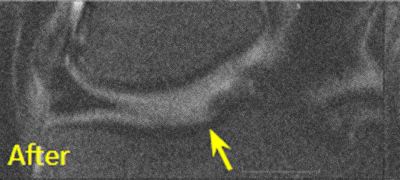
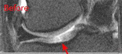
Patient 1 MRI: 51 years old
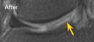
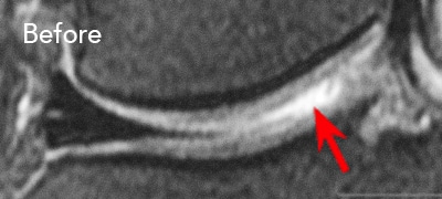
Patient 2 MRI: 46 years old
Patient 1: This individual experienced limited improvement following a microfracture procedure and was unable to resume many daily activities. A procedure using a percutaneous injection of autologous cells prepared through Regenexx lab processes was later performed. Following the procedure, the individual reported returning to routine functional activities
Patient 2: This individual underwent arthroscopic debridement, during which a 3 cm by 4 cm osteochondral defect on the medial femur was identified. Approximately 1.5 years after surgery, the individual received a percutaneous procedure involving autologous cells processed using Regenexx lab methods. Subsequent to the procedure, the individual reported resuming full functional activities.
Am I a candidate?Patient FAQs
The knee is a joint, and it is the largest one in the body. It is the point where the thigh bone (femur) connects with the shin bones (tibia and fibula). The ends of these bones are covered with a smooth, slippery tissue called cartilage, which may help reduce friction within the joint. This surface allows the bones to move against one another more easily and may help protect the joint from stress. The knee also contains two crescent-shaped pieces of fibrous cartilage called the meniscus. These structures contribute to joint stability and help distribute weight more evenly.
Over time, the knee joint is subjected to significant stress during weight-bearing activities. Like all joints, it goes through natural cycles of wear and repair, which can include cartilage thinning, the development of bone spurs, and other structural changes. In some cases, the body’s repair response can alter the joint’s shape or alignment. When this occurs, the bones may not remain properly aligned or adequately lubricated. Over time, this may lead to inflammation, pain, swelling, and stiffness, which are often associated with a condition called osteoarthritis. Contributing factors may include excess body weight, the natural aging process, or previous injury to the joint.
Arthritis is a general term used to describe inflammation of the joints. There are two primary types of arthritis:
- Osteoarthritis (OA) – also referred to as degenerative joint disease- is the most common form of arthritis. It occurs when the cartilage within a joint gradually wears away, commonly affecting the hips, knees, and spine.⁶
- Rheumatoid arthritis (RA) – is an autoimmune condition in which the immune system mistakenly targets the lining of the joints (synovium), resulting in inflammation and joint damage.⁷
Yes. Both imaging methods can detect joint degeneration, but MRI offers earlier and more detailed views. Physicians at the Rehabilitation Medicine Center of New Jersey, NY may use MRI to detect early signs of osteoarthritis and better evaluate joint health.
Although cartilage plays a key role in joint function, its loss is not typically the direct source of pain. The MRI finding most consistently linked with pain is swelling within the bone, known as bone marrow edema (BME) or a bone marrow lesion (BML). Recent research on knee pain related to arthritis has shifted focus from cartilage deterioration to the presence of this bone marrow swelling.
No, they will not regenerate large areas of cartilage; however, they may help:
- Replenish diminished cellular reserves. The body’s natural supply of cells involved in repair and maintenance tends to decline with age and the progression of arthritis.
- Support cellular recovery. Research suggests that mesenchymal stem cells may transfer healthy mitochondria to neighboring cells with damaged mitochondria, potentially restoring function in those cells.
- Address bone lesions. Emerging evidence indicates that microfractures, rather than cartilage loss, are a primary contributor to pain.
- Modulate the joint environment. Arthritic joints often contain a mix of inflammatory and catabolic substances, and cellular therapies may help shift the environment toward one that is more supportive of tissue maintenance.
Request an Appointment
References
- 4. Regenexx Patient Registry – Knee Outcomes. Lower Extremity Functional Scale (LEFS) questionnaire. Accessed on November 02, 2020.
Moseley JB, O’Malley K, Petersen NJ, Menke TJ, Brody BA, Kuykendall DH, Hollingsworth JC, Ashton CM, Wray NP. A controlled trial of arthroscopic surgery for osteoarthritis of the knee. N Engl J Med. 2002 Jul 11;347(2):81-8. doi: 10.1056/NEJMoa013259. PMID: 12110735. [Google Scholar]
Englund M, Guermazi A, Gale D, Hunter DJ, Aliabadi P, Clancy M, Felson DT. Incidental meniscal findings on knee MRI in middle-aged and elderly persons. N Engl J Med. 2008 Sep 11;359(11):1108-15. doi: 10.1056/NEJMoa0800777. PMID: 18784100. [Google Scholar]
Katz JN, Brophy RH, Chaisson CE, de Chaves L, Cole BJ, Dahm DL, Donnell-Fink LA, Guermazi A, Haas AK, Jones MH, Levy BA, Mandl LA, Martin SD, Marx RG, Miniaci A, Matava MJ, Palmisano J, Reinke EK, Richardson BE, Rome BN, Safran-Norton CE, Skoniecki DJ, Solomon DH, Smith MV, Spindler KP, Stuart MJ, Wright J, Wright RW, Losina E. Surgery versus physical therapy for a meniscal tear and osteoarthritis. N Engl J Med. 2013 May 2;368(18):1675-84. doi: 10.1056/NEJMoa1301408. Epub 2013 Mar 18. Erratum in: N Engl J Med. 2013 Aug 15;369(7):683. PMID: 23506518 [Google Scholar]
Sihvonen R, Englund M, Turkiewicz A, Järvinen TL; Finnish Degenerative Meniscal Lesion Study Group. Mechanical Symptoms and Arthroscopic Partial Meniscectomy in Patients With Degenerative Meniscus Tear: A Secondary Analysis of a Randomized Trial. Ann Intern Med. 2016 Apr 5;164(7):449-55. doi: 10.7326/M15-0899. Epub 2016 Feb 9.PMID: 26856620. [Google Scholar]
van de Graaf VA, Noorduyn JCA, Willigenburg NW, Butter IK, de Gast A, Mol BW, Saris DBF, Twisk JWR, Poolman RW; ESCAPE Research Group. Effect of Early Surgery vs Physical Therapy on Knee Function Among Patients With Nonobstructive Meniscal Tears: The ESCAPE Randomized Clinical Trial. JAMA. 2018 Oct 2;320(13):1328-1337. doi: 10.1001/jama.2018.13308. Erratum in: JAMA. 2018 Dec 4;320(21):2272-2273. Erratum in: JAMA. 2020 Mar 10;323(10):1001. PMID: 30285177. [Google Scholar]
Regenexx Patient Registry – Knee Outcomes. Numeric Pain Scale (NPS) questionnaire. Accessed on November 02, 2020.
Regenexx Patient Registry – Knee Outcomes. Single Assessment Numeric Evaluation (SANE) questionnaire. Accessed on November 02, 2020.
NIH: National Institute of Arthritis and Musculoskeletal and Skin Diseases. Accessed October 5, 2020.
Arthritis Foundation. Accessed November 02, 2020.
Xia Z, Ma P, Wu N, Su X, Chen J, Jiang C, Liu S, Chen W, Ma B, Yang X, Ma Y, Weng X, Qiu G, Huang S, Wu Z. Altered function in cartilage derived mesenchymal stem cell leads to OA-related cartilage erosion. Am J Transl Res. 2016 Feb 15;8(2):433-46. PMID: 27158337. [Google Scholar]