A Mess of a BMA Study Can Teach Us About Orthobiologic Research
One of the hallmarks of good science is trying to limit variables. That’s what makes clinical research so hard, as making sure that there are only one or two differences between one group of patients and another isn’t always easy. That gets less complex when you’re measuring the lab metrics of something like a bone marrow aspiration, a study we’ve done many times. However, a recent paper comparing different BMA techniques changed seven different variables and has my head spinning in trying to make sense of it all at 4 am. Let’s dive in.

Why Review Research Like This at 4 am?
As Chief Medical Officer for Regenexx, it’s my job to know the published literature on orthobiologics like the back of my hand. In addition, that includes not just reading the abstract, but digging deeper to see if that research can support its conclusions. So when I found this new paper on BMA techniques, I was excited. Could this have new information that our network doctors could use? However, before I got too pumped, I needed to review the methods to determine whether the authors could support their conclusions.
BMC and BMA
BMC means bone marrow concentrate, which is a procedure where the doctor takes a bone marrow aspirate (BMA) (usually from the back of the pelvis) and then uses a centrifuge to remove the stem cell containing fraction for treatment. The good news is that our group and others have published randomized controlled trials showing that high-dose, precise BMC injections can help many common orthopedic problems like knee arthritis, shoulder rotator cuff tears, and ACL tears (1-5).
The BMA Is the First Key Step
One of the biggest problems with a BMC procedure is that many physicians perform what I call “a lazy draw”. That means that rather than taking their time to draw small volumes from many spots, which we know will increase the stem cell content of the BMA, they instead draw a huge amount of volume from a single spot, which we know will dramatically reduce stem cell content (6-8). Why perform a BMC the lazy way? Because it saves time and frankly, the patient will never know the difference.
So what happens if the doctor performs a lazy draw? Then it’s garbage in/garbage out for creating BMC. This is a serious problem, as we know through multiple studies that the stem cell content of the BMC is critical for orthopedic procedure success (9-11). Meaning that the more stem cells in the BMC, the more likely it is that the procedure will be effective. Hence, this laziness seriously short-changes the patient.
So when I meet a patient who has tried a BMC “stem cell” procedure that didn’t work, the first thing I look at is the doctor’s notes on the BMA. Was it a lazy draw or a high-quality draw? There are other things to review as well, such as how the BMC was created (simple bedside machine versus lab), how the BMC was injected, etc… However, seeing a lazy draw tells me everything I need to know about the clinic.
Keeping BMA Quality High
The first step in keeping BMA quality high is counting cells. This makes sense, as you can audit the quality of the draws this way, so if a provider is using the lazy technique this will reduce the total nucleated cell count of the BMC and that provider can be identified for retraining. However, 99% of the doctors performing these draws outside of the Regenexx network do not count the cells, hence there is nobody monitoring quality.
Testing BMA Techniques
Through the years, our Colorado research team and lab have performed many different BMA studies. We’ve published some of those and other datasets we’ve kept unpublished (12, 13). For example, many years ago we tested a new device called Marrow Cellutions that was supposed to allow you to avoid concentration of the BMA, but didn’t work as advertised. This past month we tested a new marrow wash technique, which is data I may share at some point.
One of the key factors in producing solid research on the yields of different BMA techniques is following the simplest rule in science, keep your variables limited. What does that mean?
Variables 101
In medical research, we’re often testing one group against another. That works well when you test one difference or variable between the groups. In the specific case of testing BMA techniques that might look like 30 patients in group 1 where you go deep and draw 5 ml and the same 30 patients in group 2 where you stay superficial and draw the same 5 ml from the other side. However, what happens if you change too much at once? Let’s say in group 1 you go deep and change the way the trocar needle is used and draw a higher volume compared to group 2. Now if you have differences in the groups in the number of stem cells collected and you have no idea of what caused that effect. Or if you isolate some of these changes to parts of the group, you begin to get too few patients who each have the same draw conditions. That also threatens the integrity of your results because a difference seen in 5-10 patients may disappear at 20-30 patients.
Now that we understand BMAs, quality control, and variables, let’s look at a new study that tests so many different variables in so few unique patients that it’s hard to know what they found.
The New Research
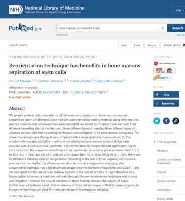
This one is out of Austria and involves 48 BMAs in 17 patients (14). The paper is very tough to follow. Basically, the problem is that they changed so many different variables at the same time that the number of patients in any single distinct group compared to any other single distinct group are very small. Let’s begin with all of the things being changed and compared in these patients:
- Orientation of the trocar: This is a single orientation of the trocar versus reorienting it while still in the marrow.
- The volume of bone marrow being drawn (big versus small).
- The centrifuge used to create BMC (Arthrex Angel vs. IMPACT)
- The type of trocar used for the BMA (Athrex vs. Argon vs. Marrow Cellutions)
- The size of the draw syringe used for the BMA (10 ml vs 30 ml)
- The location of the draw (posterior iliac crest versus anterior)
- The side of the draw (right versus left)
Yikes! Just reading this thing early in the morning makes my head spin. Let’s look at a single patient where conditions were changed so we can see if we can compare these to gather any meaningful data. This is patient 1 in group 1:
- Condition A — Single trocar orientation, big volume, Arthrex Angel centrifuge, Argon trocar, 10 ml syringe, anterior iliac crest draw site, right side
- Condition B — Single trocar orientation, big volume, Arthrex Angel centrifuge, Argon trocar, 10 ml syringe, posterior iliac crest draw site, right side
- Condition C — Single trocar orientation, big volume, Arthrex Angel centrifuge, Arthrex trocar, 30 ml syringe, anterior iliac crest draw site, left side
- Condition D — Single trocar orientation, big volume, Arthrex Angel centrifuge, Arthrex trocar, 30 ml syringe, posterior iliac crest draw site, left side
So a single patient here underwent what appears to be 3 draws on each side. In fact, the 9 patients in group 1 had 32 bone marrow punctures! This is a HUGE problem as the authors say little about whether these draws were done at the same time/local area or at different times. For example, we have found that performing many draws at the same time in the same general area can deplete the cells in that spot. That means that you may see a drop in total cells solely because you went back to the same “well” twice. We’ve also found that for older women, repeat draws can deplete cells when performed within 6 months of each other. So this places into question the results of this entire experiment.
Trying to read through the conditions for patient 1 in group 1, we see that anterior and posterior draw sites are compared in conditions A and B. If we just focus on that single difference between patients, how many patients are there that had this anterior/posterior comparison where everything else was the same? That’s 9 patients in the anterior group versus 7 of the same patients in the posterior group. This is also a big problem, as we’ve seen trends develop in 10 or 20 patients that when we looked at 30 or 40 patients, disappear. Meaning the authors compared far too few patients to draw any meaningful conclusions.
The numbers of patients available for these comparisons get far worse from here. For example, in Table 3 for patient 1, they change the volume (big versus small), the centrifuge, the trocar, the syringe, and the side. Meaning the numbers available for single variable comparisons are tiny.
Measuring CD34+?
As if this paper wasn’t a big enough absolute mess, the authors seem to have measured BMA metrics that have little meaning in the treatment of knee osteoarthritis based on what we know. For example, we know based on published and peer-reviewed research that the total nucleated cell count of BMC and CFU-f (a proxy for mesenchymal stem cell count) is important in determining the outcome (9-11,15). So did the authors measure either of these counts in the BMC? Nope. They measured CD34+ (Hematopoietic Stem Cells) and total white blood cells (Leukocytes). As far as I know, neither of these counts has ever been tied to the clinical outcome in the use of BMC to treat knee OA.
A Fancy Name?
Our authors gave this really bad study a name: SUSTEXAP (SUstainable STemcell EXtraction and APplication). That to me, given the crazy study design, seems like a bit of over-the-top hubris.
Why Did This Study Fail to Tell Us Anything New?
Universities, like this one in Austria, have a problem. They don’t do enough of these BMC cases for knee arthritis patients to have much experience or data. For example, here the authors tried to measure at least 7 different variables. How many patients would have been needed to get meaningful data in that type of study? In our clinical and research experience, at least 30-40 per group, so that’s 200-300 patients. How many clinical sites worldwide could get that size of a BMC study done in a reasonable time period (let’s say 1 year)? We could do it at our Colorado HQ site in 3-6 months. However, most universities wouldn’t treat that many BMC knee OA patients in 3-4 years.
Because of an inability to have the scale required to perform this study in a way that would have yielded new and important data, our authors went with many draws on the same 17 patients, which caused this paper to be a waste of time. I hope this serves as an impetus for universities that are just starting to get into the BMC research game to understand that you need to have a significant clinical operation developed to get any meaningful data collected.
The upshot? I began this review at about 4 am on a Saturday morning and it’s now almost 8 am. I would love to say that this was time well spent learning something new about BMAs that I could relay to our Regenexx network doctors. It was not.
___________________________________________
(1) Centeno C, Sheinkop M, Dodson E, Stemper I, Williams C, Hyzy M, Ichim T, Freeman M. A specific protocol of autologous bone marrow concentrate and platelet products versus exercise therapy for symptomatic knee osteoarthritis: a randomized controlled trial with 2 year follow-up. J Transl Med. 2018 Dec 13;16(1):355. doi: 10.1186/s12967-018-1736-8. PMID: 30545387; PMCID: PMC6293635.
(2) Hernigou P, Bouthors C, Bastard C, Flouzat Lachaniette CH, Rouard H, Dubory A. Subchondral bone or intra-articular injection of bone marrow concentrate mesenchymal stem cells in bilateral knee osteoarthritis: what better postpone knee arthroplasty at fifteen years? A randomized study. Int Orthop. 2020 Jul 2. doi: 10.1007/s00264-020-04687-7. Epub ahead of print. PMID: 32617651.
(3) Hernigou P, Delambre J, Quiennec S, Poignard A. Human bone marrow mesenchymal stem cell injection in subchondral lesions of knee osteoarthritis: a prospective randomized study versus contralateral arthroplasty at a mean fifteen year follow-up. Int Orthop. 2020 Apr 23. doi: 10.1007/s00264-020-04571-4. Epub ahead of print. PMID: 32322943.
(4) Centeno C, Lucas M, Stemoer I, Dodson E. IMAGE-GUIDED INJECTION OF ANTERIOR CRUCIATE LIGAMENT TEARS WITH AUTOLOGOUS BONE MARROW CONCENTRATE AND PLATELETS: MIDTERM ANALYSIS FROM A RANDOMIZED CONTROLLED TRIAL. Bio Ortho J Vol 3(1):e29–e39; October 5, 2021.
(5) Centeno C, Fausel Z, Stemper I, Azuike U, Dodson E. A Randomized Controlled Trial of the Treatment of Rotator Cuff Tears with Bone Marrow Concentrate and Platelet Products Compared to Exercise Therapy: A Midterm Analysis. Stem Cells Int. 2020 Jan 30;2020:5962354. doi: 10.1155/2020/5962354. PMID: 32399045; PMCID: PMC7204132.
(6) Batinić D, Marusić M, Pavletić Z, Bogdanić V, Uzarević B, Nemet D, Labar B. Relationship between differing volumes of bone marrow aspirates and their cellular composition. Bone Marrow Transplant. 1990 Aug;6(2):103-7. PMID: 2207448.
(7) Muschler GF, Boehm C, Easley K. Aspiration to obtain osteoblast progenitor cells from human bone marrow: the influence of aspiration volume. J Bone Joint Surg Am. 1997 Nov;79(11):1699-709. doi: 10.2106/00004623-199711000-00012. Erratum in: J Bone Joint Surg Am 1998 Feb;80(2):302. PMID: 9384430.
(8) Fennema EM, Renard AJ, Leusink A, van Blitterswijk CA, de Boer J. The effect of bone marrow aspiration strategy on the yield and quality of human mesenchymal stem cells. Acta Orthop. 2009 Oct;80(5):618-21. doi: 10.3109/17453670903278241. PMID: 19916699; PMCID: PMC2823327.
(9) Centeno CJ, Berger DR, Money BT, Dodson E, Urbanek CW, Steinmetz NJ. Percutaneous autologous bone marrow concentrate for knee osteoarthritis: patient-reported outcomes and progenitor cell content. Int Orthop. 2022 Aug 6. doi: 10.1007/s00264-022-05524-9. Epub ahead of print. PMID: 35932306.
(10) Pettine KA, Murphy MB, Suzuki RK, Sand TT. Percutaneous injection of autologous bone marrow concentrate cells significantly reduces lumbar discogenic pain through 12 months. Stem Cells. 2015 Jan;33(1):146-56. doi: 10.1002/stem.1845. PMID: 25187512.
(11) Hernigou P, Beaujean F. Treatment of osteonecrosis with autologous bone marrow grafting. Clin Orthop Relat Res. 2002 Dec;(405):14-23. doi: 10.1097/00003086-200212000-00003. PMID: 12461352.
(12) Schäfer R, DeBaun MR, Fleck E, Centeno CJ, Kraft D, Leibacher J, Bieback K, Seifried E, Dragoo JL. Quantitation of progenitor cell populations and growth factors after bone marrow aspirate concentration. J Transl Med. 2019 Apr 8;17(1):115. doi: 10.1186/s12967-019-1866-7. PMID: 30961655; PMCID: PMC6454687.
(13) Berger DR, Aune ET, Centeno CJ, Steinmetz NJ. Cryopreserved bone marrow aspirate concentrate as a cell source for the colony-forming unit fibroblast assay. Cytotherapy. 2020 Sep;22(9):486-493. doi: 10.1016/j.jcyt.2020.04.091. Epub 2020 Jun 19. PMID: 32565131.
(14) Pabinger C, Dammerer D, Lothaller H, Kobinia GS. Reorientation technique has benefits in bone marrow aspiration of stem cells. Sci Rep. 2022 Jul 8;12(1):11637. doi: 10.1038/s41598-022-15019-7. PMID: 35803965; PMCID: PMC9270485.
(15) Centeno CJ, Al-Sayegh H, Bashir J, Goodyear S, Freeman MD. A dose response analysis of a specific bone marrow concentrate treatment protocol for knee osteoarthritis. BMC Musculoskelet Disord. 2015 Sep 18;16:258. doi: 10.1186/s12891-015-0714-z. PMID: 26385099; PMCID: PMC4575428.

NOTE: This blog post provides general information to help the reader better understand regenerative medicine, musculoskeletal health, and related subjects. All content provided in this blog, website, or any linked materials, including text, graphics, images, patient profiles, outcomes, and information, are not intended and should not be considered or used as a substitute for medical advice, diagnosis, or treatment. Please always consult with a professional and certified healthcare provider to discuss if a treatment is right for you.
