Ortho 2.0 and a facet cyst causing a low back problem…
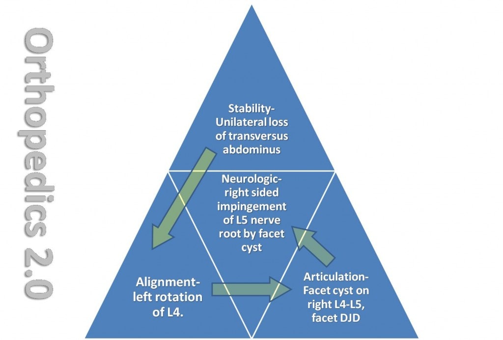
To further synthesize more concepts of why it’s important to figure out how the musculoskeletal system failed in order to strategize how it can be fixed, I present this am an interesting case of a patient from Boston who had a serious fall from a bike about three years ago. He injured his shoulder, kidney, and hip. When he was first evaluated for stem cell treatment of his hip, I was concerned about his low back. While stem cells in the hip helped the hip pain (he now now walk faster through an airport)(This patient was treated with the Regenexx-C (cultured stem cell injections), over the ensuing year he continued to develop problems in his low back and leg. He was finally was diagnosed with a cyst on his right L4-L5 facet joint, which was pressing on a nerve and giving him pain down the leg. The facet joints are small joints in the back, and sometimes arthritis of the joints can result in a cyst (just like a swollen knee joint can develop a Baker’s cyst). These cysts can press on spinal nerves, so they can be a double whammy for the patient. In Boston, he had the facet cyst treated with a steroid injection to pop the cyst. This helped some of the leg symptoims and severe nerve pain, but by the time I re-examined him recently, his back was pretty bad (unable to stand straight). This was impacting his work as a university physician. While knowing he has a facet cyst is a good strat to helping, asking the question of how he got that way is important if this is going to be successfully treated with surgical fusion of this level. His case is a good example of the ortho 2.0 concept. Consider the ortho 2.0 pyramid below, in which I’ve filled in various portions:
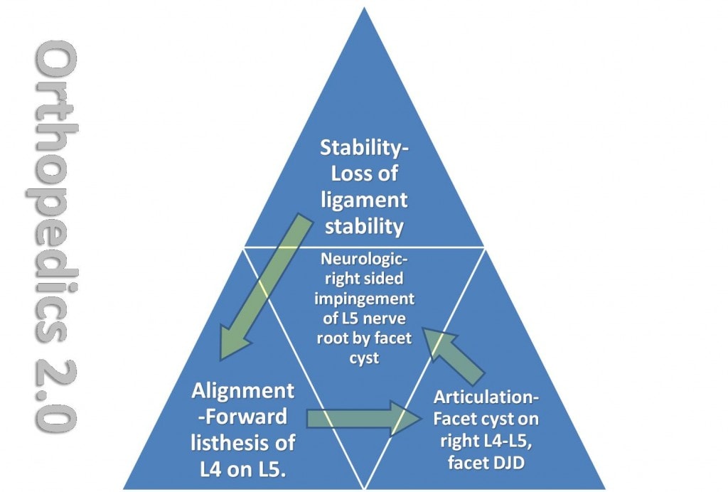
To better explain, more discussion and pictures are needed. On his flexion-extension views, it was noted that he had the L4 vertebra slipping forward on the L5 vertebra. This forward slippage was at the same level as his facet cyst. Coincidence? Likely not. The way to understand this problem involves some ligaments in the back of the spine that act as the major duct tape that help keep the spine aligned. These ligaments are the supraspinous and interspinous ligaments. The image below shows the ligaments (red lines) in the back of the spine.
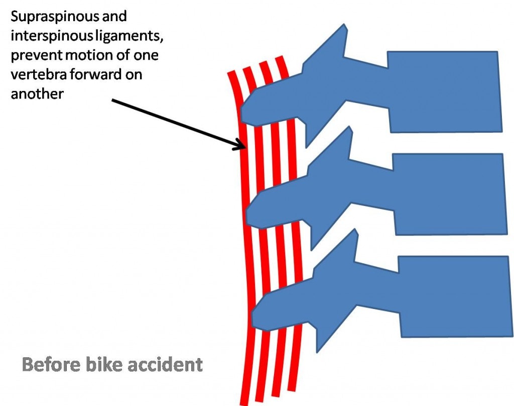
So before his bike accident, these ligaments are doing their job, helping to hold the spine in alignment. After the accident, this is what I believe happened:
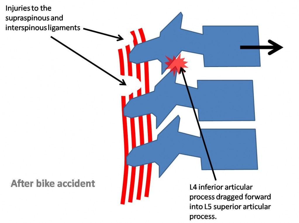
Note that after the bike crash, the injury and tearing of these ligaments allow the L4 vertebra to move forward on the L5 vertebra. This causes the facet joints (above red star, there are two at each level) to get compressed, which ultimately leads to excess wear and tear of these joints. Is there another part of this puzzle? Well an interesting observation as he lies prone on the table is below:
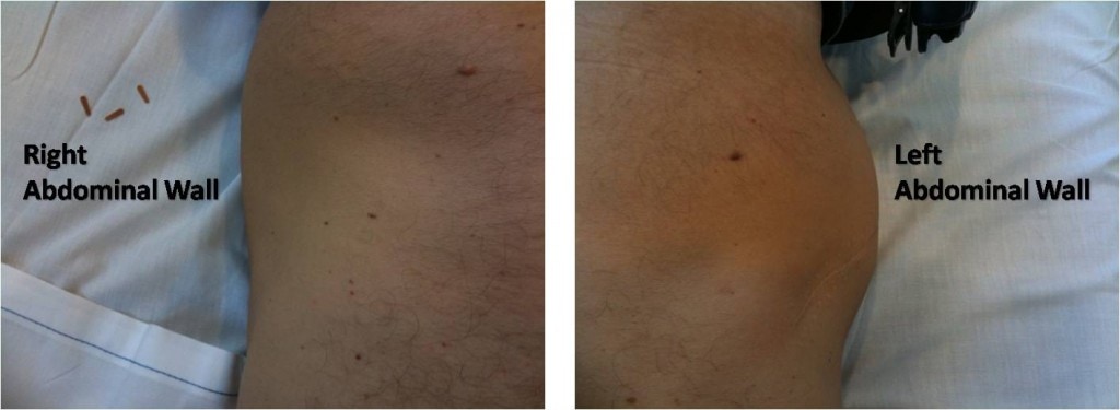
What gives with the severe bulging of left abdominal wall? Further questioning of the patient reveals that he also injured his kidney in the bike accident and had surgery on the left. The scar can be seen in the picture above. An important stabilizer of the back is the transversus abdominus. This muscle was likely cut through to get at the kidney, resulting in the muscle weakness you see above on the left side (inability to hold in the abdominal contents). This resulted in the following analysis:
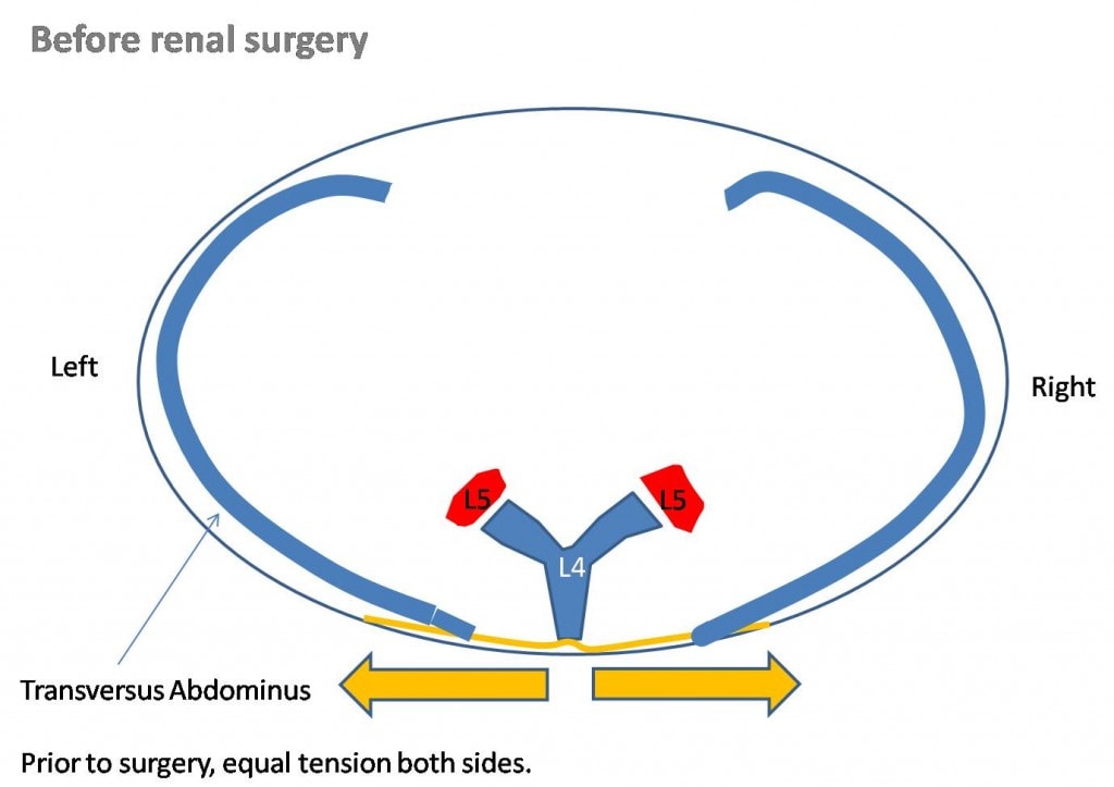
The transversus abdominus is a muscle that’s the deepest of the abdominal wall. It attaches to the thoracodorsal fascia and pulling on this muscle on both sides helps to allow the buoyancy of the abdominal contents to assist in off loading the weight of the upper body by literally floating it on the abdominal contents. It’s also a major low back stabilizer all by itself. The picture below shows that it attaches to fascia that then attaches to the back of the vertebra on both sides (spinous process). This axial view (saw you in half view) shows that if the pull is equal on both sides, this helps to keep the vertebra straight.
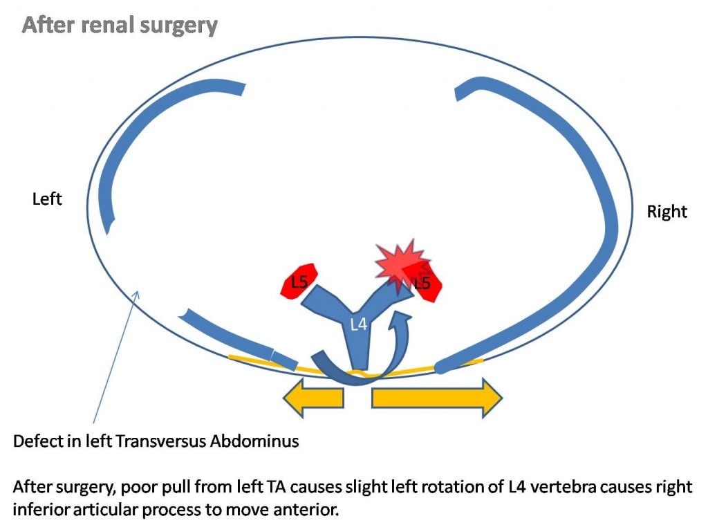
However, if we cut one side of the transversus abdominus muscle (for example to get to a damaged kidney), the forces on the vertebra will be unequal, causing it to have a slight tendency to rotate (in this case to the left). This forces on the right facet joint will increase, causing more wear and tear forces on that side.
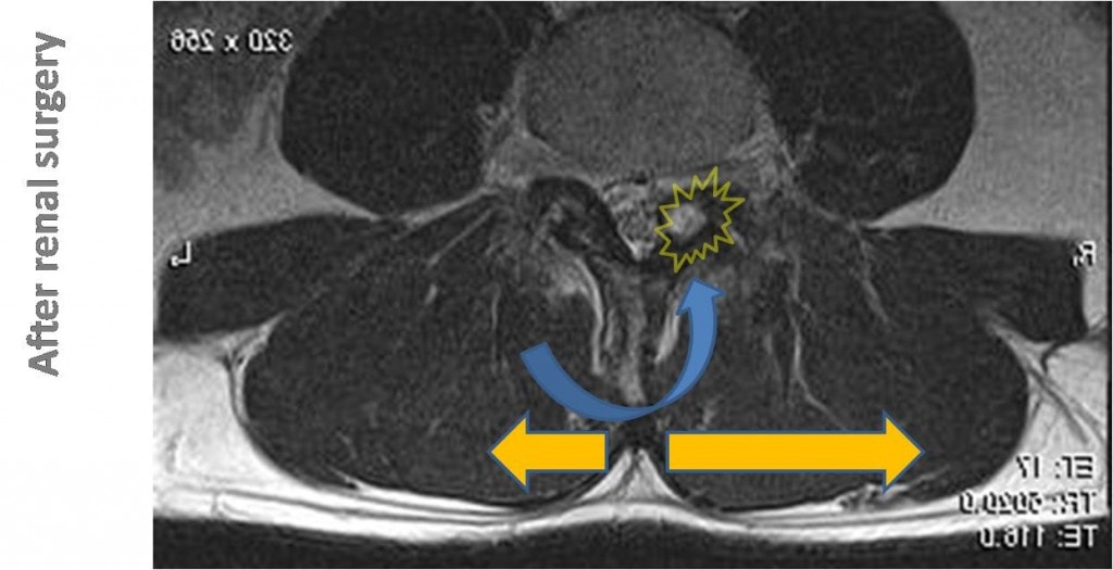
Below is an actual axial MRI image which shows the abnormal pull to the right by the transversus abdominus muscles (orange arrows) causing extra force on the right facet joint (yellow star). This is where the facet cyst is located.
So in summary, we believe that the damage to the ligaments in the back (supraspinous and interspinous ligaments) as well as this abnormal pull of one transversus abdominus over the other, have caused the facet joints to wear out. Their response on the right (the side where we would predict the most force) is to swell to try and keep up with the wear and tear. This lead to a facet cyst and then ultimately pressure on the spinal nerve. We are now designing a treatment plan for this gentleman, but I think this case illustrates the importance of piecing together all of the parts and pieces of what caused the musculosketal system to fail. Many times treating patients with musculoskeletal problems is as simple as a quick fix (in this case popping a facet cyst with a facet injection), othertimes it takes considerably more analysis.

If you have questions or comments about this blog post, please email us at [email protected]
NOTE: This blog post provides general information to help the reader better understand regenerative medicine, musculoskeletal health, and related subjects. All content provided in this blog, website, or any linked materials, including text, graphics, images, patient profiles, outcomes, and information, are not intended and should not be considered or used as a substitute for medical advice, diagnosis, or treatment. Please always consult with a professional and certified healthcare provider to discuss if a treatment is right for you.