What Are Acetabular Cysts? Exploring Cysts On The Hip

Medically Reviewed By:
The hip joint is essential for mobility and stability, making any issue affecting it a significant concern. Acetabular cysts may disrupt the function of the joint, leading to discomfort, stiffness, and limitations in movement.
While some individuals may notice only minor inconveniences, others might experience considerable pain and reduced range of motion, impacting daily activities like walking, climbing stairs, or standing for extended periods.
These challenges can profoundly affect quality of life, making it difficult to maintain an active lifestyle or carry out routine tasks comfortably. Understanding acetabular cysts, including their causes, symptoms, and available diagnostic and treatment options, provides valuable insights for managing hip health and maintaining overall well-being.
Anatomy Of The Acetabulum And Its Role In The Hip Joint
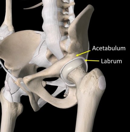
The acetabulum is a critical hip joint structure located on the pelvis. It forms the socket portion of the ball-and-socket joint, where the head of the femur (thighbone) fits securely, creating a balance between stability and flexibility. This design allows for a wide range of motion, enabling activities such as walking, running, bending, and rotating the leg.
Situated at the point where three pelvic bones—the ilium, ischium, and pubis—meet, the acetabulum features a smooth surface lined with cartilage. This cartilage serves to cushion the joint, reduce friction during movement, and evenly distribute weight across the joint. Surrounding the acetabulum is the labrum, a ring of cartilage that enhances socket depth and supports joint stability.
The acetabulum’s location and function are essential for weight-bearing activities and maintaining balance. Structural disruptions, including the formation of acetabular cysts, may impair its function, potentially resulting in pain, stiffness, and reduced mobility.

What Is An Acetabular Cyst?
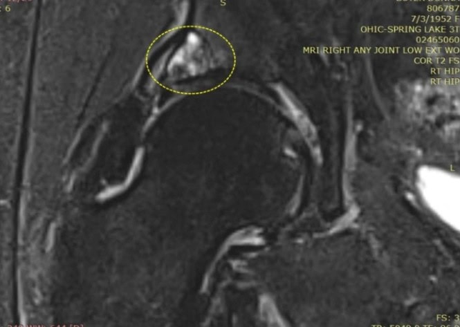
An acetabular cyst is a fluid-filled sac that forms when bone tissue in the acetabulum—the hip joint socket—degenerates or undergoes necrosis (cell death). This condition typically arises when the forces exerted on the bone exceed the body’s capacity to repair the damage. In response, the body attempts to isolate the affected area by forming a dense, protective shell around it, resulting in the formation of a spherical cyst.

The image above illustrates multiple small acetabular cysts within the hip joint, highlighted by the yellow dashed circle. The white regions represent the extent of cystic damage, demonstrating how smaller cysts may merge to form larger ones over time. This progression can compromise the acetabulum’s structure and function, potentially resulting in pain, reduced mobility, and joint instability.
Acetabular cysts often develop in conjunction with conditions like osteoarthritis, where the wear and tear of cartilage exposes the underlying bone to damage. They may also result from trauma that disrupts the distribution of forces within the hip joint.
Their presence indicates ongoing stress and degeneration, underscoring the importance of a thorough evaluation to determine and address the underlying causes. Effective management is crucial to mitigate further joint damage and restore function.
Common Causes And Risks For Hip Cyst Formation
Acetabular cysts often develop due to excessive stress on the hip joint, exceeding the bone’s natural ability to repair itself and leading to localized degeneration. Their formation frequently signals underlying joint strain or structural issues. Several causes and risk factors contribute to the development of these cysts:
Hip Trauma
Trauma to the hip joint is a significant factor in developing acetabular cysts. Injuries such as fractures, dislocations, or severe impacts can disrupt the acetabulum’s structural integrity, causing localized stress and degeneration. When the bone sustains damage that exceeds its capacity to heal, the body may form a cyst to isolate the affected area.
Repetitive microtraumas, often seen in athletes or individuals involved in high-impact activities, can also result in cumulative damage over time. This ongoing strain weakens the acetabulum, increasing the likelihood of cyst formation.
Hip Dysplasia
Hip dysplasia, a condition in which the hip socket is abnormally shallow or improperly formed, is a notable risk factor for acetabular cyst formation. This structural abnormality disrupts the natural alignment of the hip joint, resulting in uneven force distribution across the acetabulum. Over time, these imbalanced forces can weaken the bone, increasing its susceptibility to degeneration and cyst development.
The altered hip joint mechanics associated with dysplasia often accelerate cartilage wear, exposing the underlying bone to heightened stress. Cysts may form a protective response to contain the damage as the bone attempts to repair itself. These cysts can further destabilize the joint, potentially exacerbating pain, stiffness, and reduced mobility.
Degenerative Changes
Degenerative changes in the hip joint are among the most common contributors to acetabular cyst formation. Conditions such as osteoarthritis progressively wear away the protective cartilage that cushions the acetabulum. As the cartilage deteriorates, the underlying bone is subjected to increased stress and friction, fostering an environment conducive to cyst development.
These changes often progress gradually as part of the aging process, but can be accelerated by factors such as obesity, repetitive joint stress, or prior injuries. Under these conditions, the weakened bone may struggle to repair itself, prompting the formation of cysts as a protective response to contain damage and shield the surrounding tissue.
Other Risk Factors
Several additional factors can contribute to the development of acetabular cysts by either increasing stress on the hip joint or compromising its structural integrity. These risk factors often interact with underlying conditions, accelerating joint degeneration and the likelihood of cyst formation. Key risk factors include:
- Age: Aging naturally increases joint wear and tear, making cartilage loss and acetabular cyst formation more common over time.
- Genetics: A family history of conditions like hip dysplasia or osteoarthritis may predispose individuals to joint vulnerabilities that can lead to cyst development.
- High-impact activities: Repetitive or high-impact movements, such as running or jumping, place excessive stress on the hip joint, raising the risk of cyst formation.
- Obesity: Excess body weight stresses the hip joint, accelerating cartilage wear, bone degeneration, and cyst formation.
Key Indicators Of Cysts Affecting The Hip Joints
Acetabular cysts can significantly impair the function of the hip joint, often causing symptoms that interfere with daily activities. These symptoms typically emerge as cysts disrupt the joint’s structural integrity and place additional stress on surrounding tissues. Recognizing these indicators early can facilitate timely evaluation and management. Common symptoms associated with acetabular cysts include:
- Pain Or Discomfort During Prolonged Sitting Or Standing: Hip pain that worsens with extended sitting or standing may indicate stress on the joint caused by cysts.
- Limited Range of Motion In The Hip: This can make everyday movements like walking, bending, or sitting uncomfortable. It may result from joint stiffness, muscle tightness, injury, or underlying conditions like arthritis. Identifying the cause is key to effective management.
- Instability Or Weakness In The Hip Joint: This can lead to difficulty standing, walking, or maintaining balance. It may result from ligament laxity, muscle weakness, injury, or joint degeneration. Identifying the underlying cause is essential for effective management.
- Clicking Or Popping Sensations In The Hip: Known as snapping hip syndrome, may occur due to tight muscles, ligament movement, or cartilage issues. While often harmless, persistent clicking with pain may indicate an underlying condition requiring evaluation.
How A Hip Cyst Is Diagnosed
Diagnosing an acetabular cyst typically begins with a comprehensive medical evaluation, including a discussion of symptoms, medical history, and a physical examination. Physicians assess pain levels, joint mobility, and any noticeable stiffness or tenderness in the hip to determine the extent of the issue. A typical evaluation includes the following steps:
- Physical examination: A physician may observe the patient’s gait, test the joint’s range of motion, and apply pressure to specific areas to pinpoint localized pain linked to the cyst.
- Imaging tests:
- X-rays: Often the initial diagnostic tool, X-rays can reveal bone abnormalities, joint degeneration, or indications of cysts in the acetabulum.
- MRI (magnetic resonance imaging): Provides detailed images of soft tissues, cartilage, and bone, making it the gold standard for identifying cysts and assessing their extent.
- CT scans (computed tomography): Offer high-resolution views of bone structure, helping to confirm the presence and size of acetabular cysts.
- Joint fluid analysis: In some cases, fluid from the joint may be extracted and tested to rule out infections or inflammatory conditions contributing to symptoms.
Conventional Treatment Approaches
Managing acetabular cysts involves strategies to alleviate symptoms, improve joint function, and address underlying causes. Physicians often recommend conservative treatments before considering more advanced interventions. Common treatment approaches include:
- Activity Modification – Avoiding high-impact activities may help reduce stress on the hip joint.
- Physical Therapy – Strengthening exercises can improve joint stability and flexibility.
- Arthroscopic Surgery – A minimally invasive procedure to remove the cyst and insert a bone graft to relieve pain and support joint function.
- Hip Replacement – In cases where arthritis coexists with the cyst, joint replacement may be considered to help address joint damage and support mobility in select patients.
Interventional Orthobiologic Options for Acetabular Cysts
Interventional orthobiologics rely on cells and growth-factor rich plasma drawn from an individual’s blood or bone marrow. In the Regenexx network, every sample is processed in an accredited on-site laboratory rather than a single-spin bedside device. Laboratory processing allows clinicians to adjust cell concentrations from six to twenty times baseline, creating a preparation that matches the specific needs of the hip joint.
Under live imaging, the material is guided to areas of degeneration in the acetabulum. This less invasive strategy seeks to calm joint inflammation, support tissue repair, and maintain mobility without the extended recovery common after surgery.
Interventional orthobiologic procedures may include:
- Regenexx SCP Platelet-Rich Plasma: Prepared on-site for high growth-factor concentration; image-guided injection aims to reduce inflammation and support soft-tissue repair.
- Stem Cell Containing Bone Marrow Concentrate: Custom concentrated marrow cells are precisely placed into damaged hip bone to encourage cartilage stability and bone health.
Each image-guided procedure fits into a wider plan for protecting joint health and improving function over time.
Patient MRI Results And Acetabular Cyst Treatment

Donna underwent a hip bone marrow concentrate (BMC) injection procedure using her own bone marrow concentrate containing mesenchymal stem cells to address her acetabular cyst. The treatment involved targeted injections into the arthritic joint, the damaged labrum, and the acetabular bone.
For the acetabular cyst, a micro-trocar (3.5-inch, 15-gauge) was used under X-ray guidance to deliver the injection precisely into the cyst. This procedure was performed using image guidance and required a recovery period.
Outcome:
When Donna returned to Colorado a year later, she reported changes in her mobility, including a reduction in limping. A follow-up MRI showed a decrease in the cyst’s size and bone infill. Individual patient outcomes vary and depend on multiple factors including condition severity and adherence to follow-up care.
Donna reported being able to return to Pilates within weeks after her procedure. Following her procedure, Donna was able to resume Pilates within weeks. Recovery times vary depending on individual circumstances and treatment choices.
Your Next Step To Better Hip Health Starts Here
Acetabular cysts can impact mobility and daily activities. Some individuals may explore non-surgical options to support joint function and mobility. Physicians in the licensed Regenexx network provide customized interventional orthobiologic procedures that may help support joint function and mobility.
By combining targeted procedures with lifestyle modifications, such as exercise and proper movement mechanics, patients may explore options to manage acetabular cysts while maintaining an active lifestyle.
Explore non-surgical options for joint health. Consult a physician in the licensed Regenexx network to learn more.

Medically Reviewed By:
