Knee Stem Cell X-ray Results: Watch Me Grow New Cartilage in Seconds!

©Regenexx
Chiropractic offices offering dead stem cell procedures have been advertising before and after X-rays purporting to show that there is copious new cartilage regrowth in arthritic knees. To the uninitiated, these X-rays look amazing, but is this true, or like the idea that these are actual stem cell injections, is it a scam? Well, I set out this week to find out by x-raying my own knee, and let me show you how I created loads of new cartilage in seconds!
Chiro Stem Cell Scams Aplenty
The biggest issue we see with chiro, acupuncture, and naturopathy offices offering “stem cell” injection procedures is that they typically use midlevel providers to inject dead cells at top dollar. They usually use FDA-registered tissue products derived from amniotic or cord blood sources that are regulated to be nonviable, and they often tell patients that there are millions of viable and young stem cells in these shots. For info on this bait-and-switch scam, see my video below:
Understanding How X-ray Works
X-ray works like a beam of light passing through tissue. The hard stuff in the way (bones), creates a shadow on the film (or these days on the computer sensor). Hence, if you take your cell phone light and shine it at your hand and look at the shadow it casts on the desk, that’s basically the same setup. Now if you alter the position of the light source just a tiny bit, the shadow on the desk can change quite dramatically. Hence, small changes in the angle of the X-ray can mean big differences in how the X-ray looks.
My Experiment This Morning
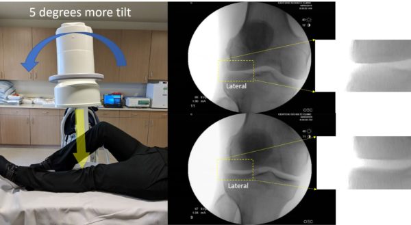
For the sake of science, I got on the table and had my left knee x-rayed. My images are to the right, and that’s the rad tech modeling how she took the images. As you can see by the top right image, the width (height) of the joint space on the lateral side of the knee looks less than the medial side. I surely have knee arthritis! However, the tech then tilted the beam just an imperceptible five degrees and that image is below right. All of my arthritis went away!
Obviously, I didn’t regrow a bunch of new cartilage on the lateral side of my knee in the seconds it took to adjust the X-ray beam and take a new image. This is merely a sleight of hand of X-ray. Change the angle of the beam slightly while x-raying a knee and one compartment can appear to have grown cartilage or have arthritis.
Where Are The MRI Before and Afters?
Early on, we published a slew of MRIs that did show some stem cell injection-related cartilage and meniscus regeneration in selected patients with less severe arthritis. First, we used real stem cells rather than dead-cell amniotic or cord blood tissue. Second, even then, we used the same state-of-the-art 3.0 Tesla scanner, with the same exact setting on both images, with the MRI taken close to the same time of day. Why? While finding the same spot on MRI is more reliable than X-ray, if the patient is lined up differently in the scanner, the MRI has lower resolution, or the settings are different, tissues can appear different even when they’re actually the same. In addition, even the time of day can impact the hydration status of cartilage and spinal discs. As a result, these tissues are generally taller in the morning and lose height due to water being pushed out by gravity during the day.
What does a real imaging before and after a stem cell treatment look like? The image below is of a patient who had her knee treated by us with culture-expanded stem cells and then had an MRI using the same settings and on the same scanner. This is a T2 map, looking at cartilage quality as well:
What Type of Cartilage Regrowth Is Possible in Real Stem Cell Treatments?
For knees with less cartilage loss (mild to moderate arthritis), cartilage regeneration with stem cells may be possible on a case-by-case basis. Below is my video of all of the great before and after MRI results we have seen through the years by using real stem cells and placing them in precisely the exact right location with imaging guidance:
However, for knees (and other joints) with more severe arthritis (more advanced moderate to severe), we don’t generally see cartilage regrowth. If you want to see how stem cell injections help symptoms and improve function without regrowing cartilage, I discuss that in more detail at this link.
Protecting Your Pocketbook at a Chiro or Alternative Medicine “Stem Cell” Seminar
So now you know about the before and after X-ray sleight of hand! However, if you attended a “stem cell” seminar given by a chiropractor, acupuncturist, or naturopath, what else do you need to look out for? To find out, watch my video below:
The upshot? As you can see, these before and after X-rays are a scam just like the dead stem cells used in these offices. So attend a local fake stem cell seminar and give them heck about why they’re not showing before and after MRIs growing cartilage! Also, mention that the way they “regrew” cartilage with dead stem cells was by merely tilting the beam a little!

If you have questions or comments about this blog post, please email us at [email protected]
NOTE: This blog post provides general information to help the reader better understand regenerative medicine, musculoskeletal health, and related subjects. All content provided in this blog, website, or any linked materials, including text, graphics, images, patient profiles, outcomes, and information, are not intended and should not be considered or used as a substitute for medical advice, diagnosis, or treatment. Please always consult with a professional and certified healthcare provider to discuss if a treatment is right for you.
